. 2009 May 15;69(10):4167-74.
doi: 10.1158/0008-5472.CAN-08-4859. Epub 2009 May 12.

The mechanical rigidity of the extracellular matrix regulates the structure, motility, and proliferation of glioma cells

Affiliations

The mechanical rigidity of the extracellular matrix regulates the structure, motility, and proliferation of glioma cells

Theresa A Ulrich et al. Cancer Res. .

Abstract

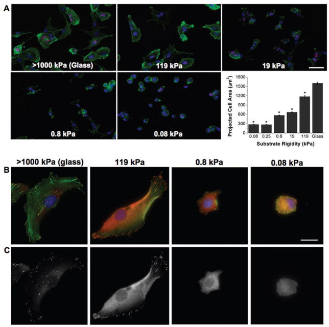
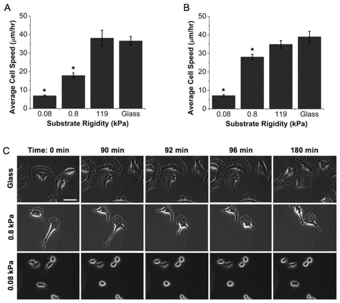
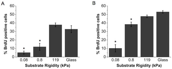
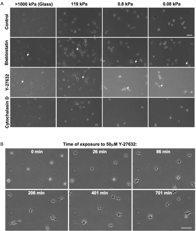
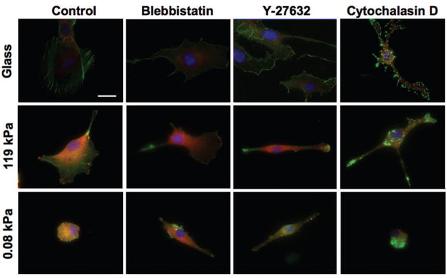
Glioblastoma multiforme (GBM) is a malignant astrocytoma of the central nervous system associated with a median survival time of 15 months, even with aggressive therapy. This rapid progression is due in part to diffuse infiltration of single tumor cells into the brain parenchyma, which is thought to involve aberrant interactions between tumor cells and the extracellular matrix (ECM). Here, we test the hypothesis that mechanical cues from the ECM contribute to key tumor cell properties relevant to invasion. We cultured a series of glioma cell lines (U373-MG, U87-MG, U251-MG, SNB19, C6) on fibronectin-coated polymeric ECM substrates of defined mechanical rigidity and investigated the role of ECM rigidity in regulating tumor cell structure, migration, and proliferation. On highly rigid ECMs, tumor cells spread extensively, form prominent stress fibers and mature focal adhesions, and migrate rapidly. As ECM rigidity is lowered to values comparable with normal brain tissue, tumor cells appear rounded and fail to productively migrate. Remarkably, cell proliferation is also strongly regulated by ECM rigidity, with cells dividing much more rapidly on rigid than on compliant ECMs. Pharmacologic inhibition of nonmuscle myosin II-based contractility blunts this rigidity-sensitivity and rescues cell motility on highly compliant substrates. Collectively, our results provide support for a novel model in which ECM rigidity provides a transformative, microenvironmental cue that acts through actomyosin contractility to regulate the invasive properties of GBM tumor cells.


Conflict of interest statement

Conflicts of Interest: None.

Figures

Figure 1
Figure 1. ECM rigidity alters glioma cell morphology and cytoskeletal organization
(A) Rigidity-dependent changes in cell structure. U373-MG cells cultured on fibronectin-conjugated glass and polyacrylamide gels over a range of stiffnesses were stained for F-actin (green), nuclear DNA (blue) and the nuclear antigen Ki67 (red). Note that a subset of cells on all substrates stained positive for Ki67. Bar is 50 μm. (*p < 0.01 with respect to glass; n > 450 cells for each condition) (B) High-magnification imaging of cytoskeletal and adhesive structures. U87-MG cells were stained for F-actin (green), nuclear DNA (blue), and the focal adhesion protein vinculin (red). Bar is 25 μm. (C) Isolated view of vinculin signal only, showing structure and distributions of cell-ECM adhesions.
Figure 2
Figure 2. ECM rigidity regulates glioma cell motility
Effect of ECM rigidity on the random migration speed of (A) U373-MG and (B) U87-MG cells. Results represent the average migration rate from at least 15 cells per condition over 6–12 hours. Qualitatively similar dependences of migration speed on substrate stiffness were observed for SNB19, U251-MG, and C6 cells. (*p < 0.01 with respect to glass) (C) High-magnification imaging of U373-MG cell migration on ECMs of varying rigidity over both long time scales (columns 1–2 and 4–5) and short time scales (columns 2–4). Cells on glass (top row) migrate quickly, smoothly, and with broad, stable lamellipodia. Cells on 0.8 kPa ECMs (middle row) form smaller, less stable lamellipodia, and migrate in a “stick-slip” fashion, in which the cell thins and extends as it advances and then abruptly contracts as adhesions at the trailing edge rupture. Cells on 0.08 kPa ECMs (bottom row) continuously extend thin filopodia and fail to productively migrate. Bar is 50 μm.
Figure 3
Figure 3. ECM rigidity regulates glioma cell proliferation
Effect of ECM rigidity on proliferation of (A) U373-MG and (B) U87-MG cells. Results represent quantification of n > 325 cells in at least 8 fields of view per substrate for at least five substrates per condition, where the percentage of dividing cells was determined as the average percentage of cells staining positive for BrdU incorporation. (*p < 0.01 with respect to glass).
Figure 4
Figure 4. Pharmacologic inhibition of cytoskeletal contractility reduces stiffness-dependent differences in cell morphology
(A) U87-MG cells cultured on fibronectin-conjugated glass and polyacrylamide substrates in either the absence of drug (control) or 24 hours after addition of 25 μM blebbistatin, 10 μM Y-27632, or 1 μM cytochalasin D. Cells began extending actin-rich processes (arrows) within an hour after addition of Y-27632 or blebbistatin. Cytochalasin D induced stellation and rounding of cells on stiff substrates but had no appreciable effect on the morphology or migration of cells on the softest substrates. Bar is 100 μm. (B) U373-MG cells cultured on 0.08 kPa fibronectin-conjugated polyacrylamide substrates showed enhanced spreading and migration with addition of 50 μM Y-27632. Bar is 100 μm.
Figure 5
Figure 5. Pharmacologic inhibition of cell tension reduces stiffness-dependent differences in cytoskeletal and adhesive architecture
U87-MG cells cultured on fibronectin-coated glass and polyacrylamide substrates were treated with 25 μM blebbistatin, 10 μM Y-27632, or 1 μM cytochalasin D for 12–24 hours before fixation and staining for nuclear DNA (blue), F-actin (green), and vinculin (red). In all cases, the number and size of vinculin-positive focal adhesions was reduced with inhibition of cell tension. Blebbistatin and Y-27632 both induced cell spreading on the softest substrates, whereas cytochalasin D induced collapse of the actin cytoskeleton. Bar is 25 μm.

Similar articles

Cited by

References

    1. Nakada M, Nakada S, Demuth T, Tran NL, Hoelzinger DB, Berens ME. Molecular targets of glioma invasion. Cell Mol Life Sci. 2007;64:458–78. - PMC - PubMed
    1. Beadle C, Assanah MC, Monzo P, Vallee R, Rosenfeld SS, Canoll P. The Role of Myosin II in Glioma Invasion of the Brain. Mol Biol Cell. 2008;19:3357–68. - PMC - PubMed
    1. Manning TJ, Jr, Parker JC, Sontheimer H. Role of lysophosphatidic acid and rho in glioma cell motility. Cell Motil Cytoskeleton. 2000;45:185–99. - PubMed
    1. Salhia B, Rutten F, Nakada M, et al. Inhibition of Rho-kinase affects astrocytoma morphology, motility, and invasion through activation of Rac1. Cancer Res. 2005;65:8792–800. - PubMed
    1. Mahesparan R, Tysnes BB, Read TA, Enger PO, Bjerkvig R, Lund-Johansen M. Extracellular matrix-induced cell migration from glioblastoma biopsy specimens in vitro. Acta Neuropathol. 1999;97:231–9. - PubMed

Publication types