. 2009 May 15;69(10):4125-33.
doi: 10.1158/0008-5472.CAN-08-4402. Epub 2009 May 12.

Haploinsufficiency of Krüppel-like factor 5 rescues the tumor-initiating effect of the Apc(Min) mutation in the intestine

Affiliations

Haploinsufficiency of Krüppel-like factor 5 rescues the tumor-initiating effect of the Apc(Min) mutation in the intestine

Beth B McConnell et al. Cancer Res. .

Abstract

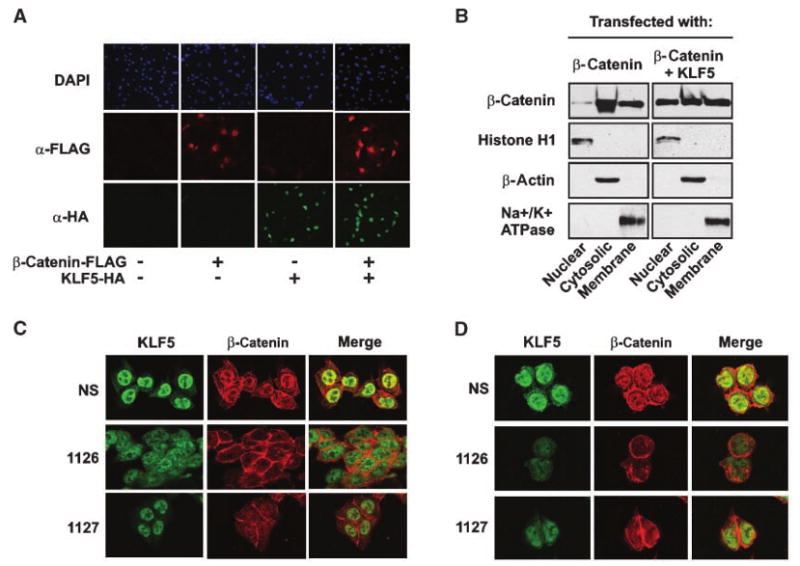
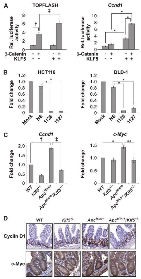
Inactivation of the tumor suppressor adenomatous polyposis coli, with the resultant activation of beta-catenin, is the initiating event in the development of a majority of colorectal cancers. Krüppel-like factor 5 (KLF5), a proproliferative transcription factor, is highly expressed in the proliferating intestinal crypt epithelial cells. To determine whether KLF5 contributes to intestinal adenoma formation, we examined tumor burdens in Apc(Min/+) mice and Apc(Min/+)/Klf5(+/-) mice. Compared with Apc(Min/+) mice, Apc(Min/+)/Klf5(+/-) mice had a 96% reduction in the number of intestinal adenomas. Reduced tumorigenicity in the Apc(Min/+)/Klf5(+/-) mice correlated with reduced levels and nuclear localization of beta-catenin as well as reduced expression of two beta-catenin targets, cyclin D1 and c-Myc. In vitro studies revealed a physical interaction between KLF5 and beta-catenin that enhanced the nuclear localization and transcriptional activity of beta-catenin. Thus, KLF5 is necessary for the tumor-initiating activity of beta-catenin during intestinal adenoma formation in Apc(Min/+) mice, and reduced expression of KLF5 offsets the tumor-initiating activity of the Apc(Min) mutation by reducing the nuclear localization and activity of beta-catenin.


Conflict of interest statement

Disclosure of Potential Conflicts of Interest: The authors have no potential conflicts of interest.

Figures

Figure 1
Figure 1
Characterization of tumor incidence and tumor burden in the ApcMin/+ and ApcMin/+/Klf5+/− mice. Mice at age 16 to 18 wk or age 28 wk were examined macroscopically for adenomas in the small intestine. A, graph of percentage of mice with small intestinal adenomas at age 16 to 18 wk (n = 22). B, scatter plot of the number of adenomas per mouse in the small intestine; ▲, ApcMin/+; formula image
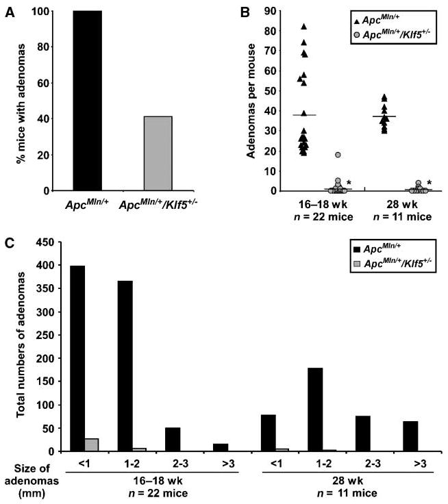
, ApcMin/+/Klf5+/−. Black horizontal lines, average number of adenomas for each genotype (*, P < 0.001; 16–18 wk, n = 22; 28 wk, n = 11; Wilcoxon-Mann-Whitney test). C, graph of total numbers of adenomas from mice screened in B, separated by size of adenomas.
Figure 2
Figure 2
Levels and localization of KLF5 and β-catenin in the small intestine of WT, Klf5+/−, ApcMin/+, and ApcMin/+/Klf5+/− mice. A, KLF5 levels in the small intestine of 16-wk-old mice were determined by immunofluorescence staining of KLF5 in normal-appearing tissues. B, quantification of KLF5 immunofluorescence intensities. Columns, mean value of four mice per group; bars, SE. Average intensities were determined from at least 50 cells per mouse. Results are shown as KLF5 fluorescence intensity per cell relative to intensities in WT mice (*, P = 0.015; **, P = 0.016; Tukey's test). C, immunohistochemical staining of β-catenin in small intestinal tissues from 16-wk-old mice. Bottom row, enlargements of the corresponding panels above. Arrows, nuclear staining. D, Western blot analysis of protein lysates from normal-appearing jejunum of 16-wk-old mice.
Figure 3
Figure 3
Interaction of KLF5 and β-catenin. A, COS-1 cells were transfected as indicated, and lysates were subjected to immunoprecipitation and Western blotting with tagged antibodies to HA (for KLF5) and FLAG (for β-catenin). B, lysates from DLD-1 cells were immunoprecipitated (IP) with either a polyclonal anti-KLF5 antibody or preimmune serum as a negative control and examined for coprecipitation of β-catenin. C, COS-1 cells were transfected as indicated and stained for immunofluorescence with antibodies derived against FLAG (red) and HA (green) to determine colocalization. D, COS-1 cells were transfected with β-catenin-FLAG alone and stained for localization of endogenous KLF5 (green) and FLAG-tagged β-catenin (red). White arrow, cell expressing ectopic β-catenin; orange arrow, cell not expressing ectopic β-catenin.
Figure 4
Figure 4
Alterations in KLF5 expression affect β-catenin nuclear localization. A, COS-1 cells were transfected as indicated and examined by immunofluorescence for nuclear β-catenin (red); KLF5 (green). Cells were counterstained with Hoechst 33528 (blue) to visualize nuclei. B, COS-1 cells were transfected with either β-catenin or β-catenin and KLF5 and fractionated into nuclear, cytosolic, and membranous components. Lysates were blotted for histone H1 (nuclear), β-actin (cytosolic), and Na+/K+ATPase (membranous) to confirm fractionation of compartments. C, HCT116 colon cancer cells were transfected with nonspecific siRNA or two distinct KLF5 siRNAs (1126 or 1127). Seventy-two hours after transfection, cells were stained by immunofluorescence for expression and localization of KLF5 (green) and β-catenin (red). D, repeat of experiment in C using DLD-1 colon cancer cells.
Figure 5
Figure 5
Changes in KLF5 expression affect activation of β-catenin transcriptional targets. A, luciferase assays in COS-1 cells transfected with expression constructs as indicated and measured for activity of TOPFLASH and Ccnd1 promoters. Rel. luciferase activity, relative luciferase activity. Coexpression of β-catenin and KLF5 produced significant synergistic effects for Ccnd1 (F [1,20] = 74.0; P < 0.001). Comparison of means (Tukey's procedure): for TOPFLASH: †, P = 0.003; ‡, P = 0.006; n = 3 per group; for Ccnd1: *, P < 0.001; n = 6 per group. B, luciferase assays in HCT116 and DLD-1 colon cancer cells transfected with indicated siRNAs. (*, P < 0.001; n = 3 per group). C, quantitative RT-PCR analysis of Ccnd1 and c-Myc mRNA levels using RNA from distal jejunum. Results were normalized to β-actin levels. For Ccnd1: †, P = 0.009; ‡, P < 0.001 0.0; n = 3; for c-Myc: *, P = 0.002; **, P = 0.009; n = 3. D, immunohistochemical staining of cyclin D1 and c-Myc in jejunum from mice at age 16 wk.
Figure 6
Figure 6
Expression of KLF5 and β-catenin in intestinal adenomas of ApcMin/+ mice. Adenomas isolated from ApcMin/+ mice at age 16 wk were stained for localization of KLF5 and β-catenin. A, representative immunohistochemical staining of KLF5 and β-catenin in ApcMin/+ adenomas. B, ApcMin/+ adenomas were stained for KLF5 (green) and β-catenin (red) by immunofluorescence to determine colocalization. Nuclei are also shown to indicate total cells.

Similar articles

Cited by

References

    1. Pinto D, Clevers H. Wnt, stem cells and cancer in the intestine. Biol Cell. 2005;97:185–96. - PubMed
    1. Kinzler KW, Vogelstein B. Lessons from hereditary colorectal cancer. Cell. 1996;87:159–70. - PubMed
    1. Moser AR, Pitot HC, Dove WF. A dominant mutation that predisposes to multiple intestinal neoplasia in the mouse. Science. 1990;247:322–4. - PubMed
    1. Su LK, Kinzler KW, Vogelstein B, et al. Multiple intestinal neoplasia caused by a mutation in the murine homolog of the APC gene. Science. 1992;256:668–70. - PubMed
    1. Fodde R. The APC gene in colorectal cancer. Eur J Cancer. 2002;38:867–71. - PubMed

Publication types

MeSH terms