. 2009 May;21(5):1373-93.
doi: 10.1105/tpc.109.065482. Epub 2009 May 12.

The NGATHA distal organ development genes are essential for style specification in Arabidopsis

Affiliations

The NGATHA distal organ development genes are essential for style specification in Arabidopsis

John Paul Alvarez et al. Plant Cell. 2009 May.

Abstract

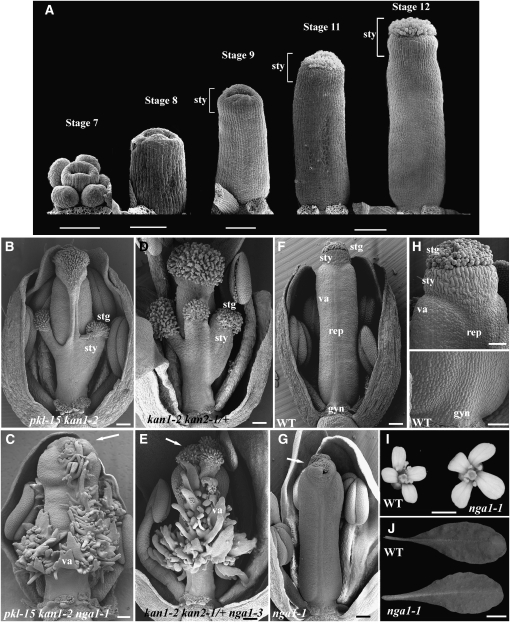
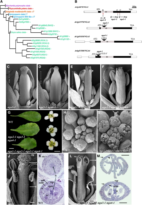
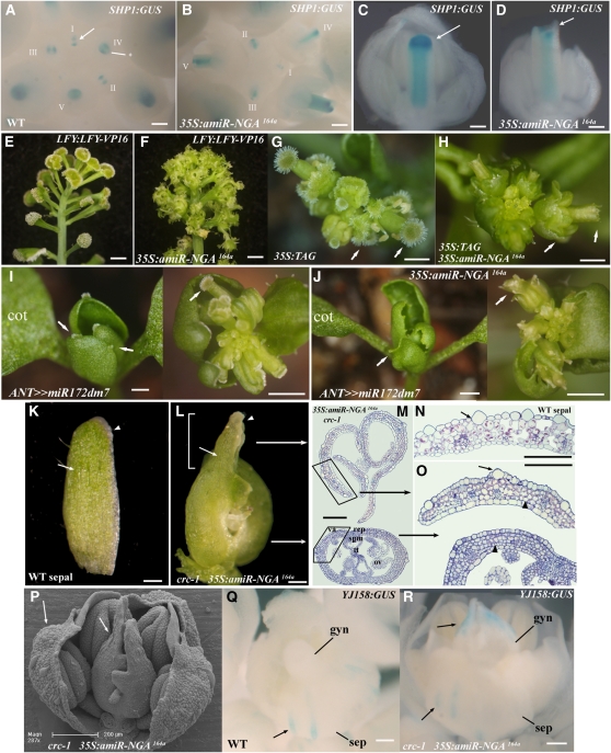
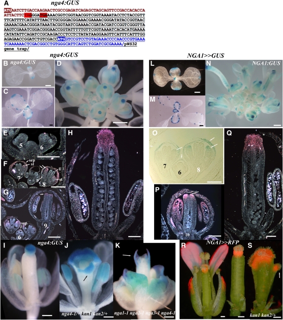
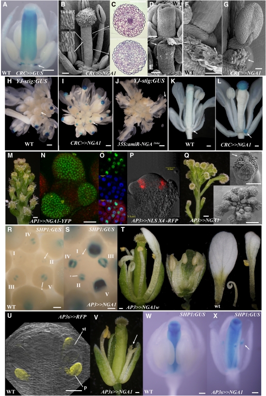
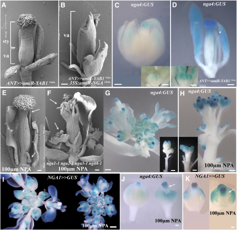
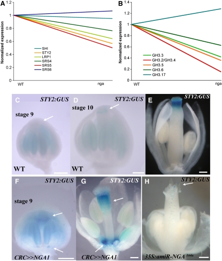
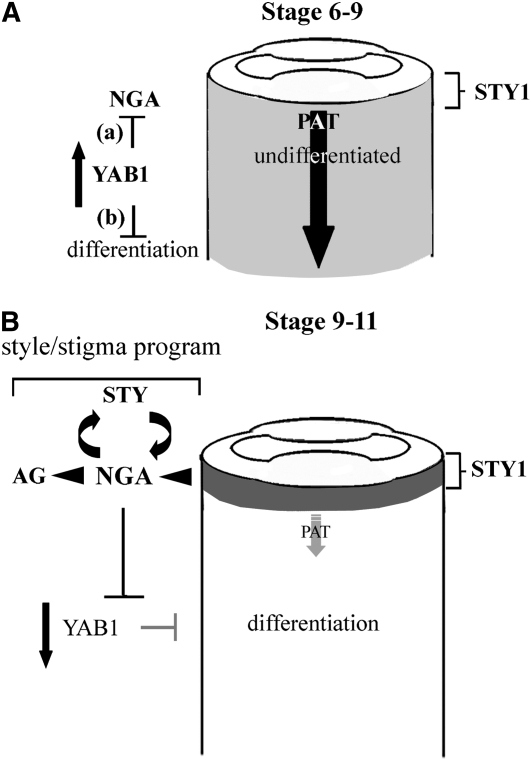
Floral organ identities are specified by a few transcription factors that act as master regulators. Subsequently, specification of organ axes programs the distribution of distinct tissue types within the organs that themselves develop unique identities. The C-class, AGAMOUS-clade MADS box genes are primary promoters of the gynoecium, which is divided into a distal style and a subtending ovary along the apical-basal axis. We show that members of a clade of B3 domain transcription factors, NGATHA1 (NGA1) to NGA4, are expressed distally in all lateral organs, and all four have a redundant and essential role in style development. Loss of all four genes results in gynoecia where style is replaced by valve-like projections and a reduction in style-specific SHATTERPROOF1 (SHP1) expression. In agreement, floral misexpression of NGA1 promotes ectopic style and SHP1 expression. STYLISH1, an auxin biosynthesis inducer, conditionally activated NGA genes, which in turn promoted distal expression of other STY genes in a putative positive feedback loop. Inhibited auxin transport or lack of YABBY1 gene activities resulted in a basally expanded style domain and broader expression of NGA genes. We speculate that early gynoecium factors delimit NGA gene response to an auxin-based signal, elicited by STY gene activity, to restrict the activation of style program to a late and distal carpel domain.


Figures

Figure 1.
Figure 1.
nga1-1 Mutant Phenotypes. (A) Scanning electron micrographs of Arabidopsis wild-type gynoecium development in progressive stages. The style becomes evident during stage 9 of flower development. All gynoecia are orientated medially. Floral stages after Smyth et al. (1990). (B) to (H) Scanning electron micrographs of medially orientated mature flowers with some outer floral organs removed to reveal the gynoecium. (B) pkl-15 kan1-2 gynoecium with basal stigma and style tissue extensions. (C) pkl-15 kan1-2 nga1-1 gynoecium with external valve and placenta outgrowths extending from the medial and basal positions of the ovary and a reduced style. (D) kan1-1 kan2-1/+ gynoecium with basal and medial style and stigmatic tissues extensions. (E) kan1-1 kan2-1/+ nga1-3 gynoecium revealing valve-like tissue as in (C). (F) Wild-type flower showing the proximal-distal elements in the gynoecium and bilateral symmetry of the ovary. (G) nga1-1 gynoecium showing disruptions in style growth. (H) Magnified view of the distal part of the wild-type gynoecium including the stigma and style (top) and basal region including the gynophore (bottom). (I) Mature wild-type and nga1-1 flowers. Note the larger petals of nga1-1 mutants. (J) Wild-type and nga1-1 leaves. nga1-1 leaves are more serrated. Arrows mark disrupted style fusion and reduced stigmatic papillae. stg, stigmatic papillae; sty, style tissue; va, valve; rep, replum; gyn, gynophore. Bars = 100 μm in (A) (stage 11 and 12) and 50 μm for stages 7 to 9, 100 μm in (B) to (G), 50 μm in (H), 1 mm in (I), and 1 cm in (J).
Figure 2.
Figure 2.
Redundant Genetic Interactions among Members of the Monophyletic NGA Clade. (A) Cladogram showing phylogenetic relationships of the RAV clade of B3 transcription factors in land plants (angiosperms,green; gymnosperms, blue; lycophyte, orange; moss, red; liverwort, purple). A complete cladogram with detailed analysis is presented in Supplemental Figure 2 online. Some members have an amino AP2 domain (+), while other members lack this domain (−). That the Physcomitrella and at least some of the Selaginella genes possess an AP2 domain suggests that it has been lost in some clades of flowering plant genes. (B) A scheme of the NGA genes. Rectangles are exons and lines are introns. Filled rectangles denote the coding region for which the start and stop codons, the ethyl methanesulfonate-generated alleles, the positions of insertions, and the highly conserved 120–amino acid B3 domains are marked. (C) to (F) Gynoecium phenotypes of lines with a progressive reduction in NGA levels. Genotypes are as labeled. (G) Leaf and flower phenotypes of nga2-1 nga3-1 nga4-1 triple and nga quadruple mutant plants relative to the wild type. The mature sixth leaf was taken in each case. (H) and (I) Scanning electron micrographs of wild-type (H) and nga quadruple mutant (I) inflorescences from an aerial perspective. (J) Scanning electron micrograph of a medial view of a wild-type gynoecium at maturity. (K) Light microscope sections through the distal style (top) and basal ovary (bottom) of a mature wild-type gynoecium taken at approximately the locations demarked by the lines in (J). The radially elongated adaxial epidermal and longitudinally elongated subepidermal cells are unique to the valve. (L) Scanning electron micrograph of a medial view of a mature nga quadruple mutant gynoecium where the style and stigma are absent and replaced by outgrowths with valve identity. (M) Light microscope sections though the distal (top) and central ovary (bottom) regions of a mature nga quadruple mutant gynoecium. The approximate, relative locations of the sections are marked by white arrows in (L). The outgrowths of tissue in the distal region have a valve identity with the typical adaxial epidermal and subepidermal cell layers (arrowhead) (K). The ovary (bottom) exhibits an apparently wild-type structure. stg, stigmatic papillae; sty, style tissue; va, valve; ade, adaxial epidermis; sade, subadaxial epidermis; rep, replum; gyn, gynophore; tt, transmitting tissue; ov, ovule; spm, septum. Bar are as marked in (C) to (F), (H), and (I). Bars = 1 cm in (G) for leaves and 1 mm for petals, and 100 μm in (J) to (M).
Figure 3.
Figure 3.
NGA Gene Activity Is Necessary for Normal and Ectopic Style Tissues. (A) to (D) Histochemical localization of SHP1:GUS in developing wild-type and 35S:amiR-NGA164a gynoecia. (A) and (B) Aerial view of wild-type (A) and 35S:amiR-NGA164a (B) inflorescences showing SHP1:GUS staining at the tips of developing gynoecia. Developmentally consecutive flowers are labeled using roman numerals. In the wild type, SHP1:GUS expression is initiated in the medial ridge, the progenitors of the placenta and septum (arrow), and later expands to the lateral and medial regions of the initiating style (asterisk). (C) Medial view of a stage 10-11 wild-type gynoecium showing intense SHP1:GUS staining in the lateral domain of the developing style (arrow). (D) Medial view of a stage 10-11 35S:amiR-NGA164a gynoecium. SHP1:GUS expression is absent from the equivalent domain (arrow). (E) and (F) Side view of inflorescences of plants homozygous for the LFY:LFY-VP16 transgene alone (E) and hemizygous for 35S:amiR-NGA164a (F). (E) LFY:LFY-VP16 flowers are composed of carpel tissue topped by enlarged stigmas. (F) 35S:amiR-NGA164a LFY:LFY-VP16 flowers lack style and stigma. (G) and (H) Aerial view of inflorescences of plants overexpressing the tomato AGAMOUS (TAG) gene alone (G) and together with 35S:amiR-NGA164a (H). (G) In 35S:TAG flowers, the sepals display extensive style and stigma tissue formation (arrow). (H) In 35S:TAG 35S:amiR-NGA164a plants, the ectopic and normal style and stigma tissues are lost (arrows). (I) and (J) Aerial views of plants overexpressing a modified version of miR172d, miR172dm7 (I) and miR172dm7 together with amiR-NGA164a (J). (I) ANT>>miR-172dm7 plants produce small leaves that are topped by stigmatic papillae (left; arrows) and flowers typically composed entirely of carpels (right; arrow). (J) ANT>>miR-172dm7 35S:amiR-NGA164a plants lacking ectopic and normal style (arrows). (K) to (R) Sepal-like identity in carpel tips lacking both CRC and NGA gene activities. (K) A wild-type sepal. Note the longitudinally elongate epidermal cells (arrow) and the pale band of marginal cells (arrowhead). (L) Gynoecium from 35S:amiR-NGA164a crc-1 flower. The distal regions of the carpels (bracketed) include large longitudinally elongated epidermal cells (arrow) and marginal band of pale cells (arrowhead) not found in normal carpels. (M) Sections though the upper and lower 35S:amiR-NGA164a crc-1 gynoecium in positions represented by the two arrows in (L). The upper gynoecium lacks carpel tissues in contrast with the base where tissues similar to those of a wild-type ovary are found. The lamina tissue in the upper gynoecium has features of both sepal and valve detailed in (N). (N) Section through a wild-type sepal. The sepal has distinct large longitudinally elongated cells at the abaxial epidermis (arrow). (O) Magnified image of the upper and lower 35S:amiR-NGA164a crc-1 gynoecium (boxed in [M]) with sepal-like cells at the upper domain (arrow). Mutant upper valves also lack the characteristic adaxial subepidermal cell layer of carpels (arrowhead). (P) Scanning electron micrograph of a 35S:amiR-NGA164a crc-1 flower with a sepal, petals, and stamens removed. Large abaxial epidermal cells can be observed in the upper carpel domain and in the sepals (arrows). (Q) Staining of YJ-158:GUS is limited to the long abaxial epidermal cells of wild-type sepals (arrow) and is not observed at any stage in the wild-type gynoecium. (R) In 35S:amiR-NGA164a crc-1 gynoecia, staining from YJ-158:GUS is also found in the long epidermal sepal-like cells in the upper gynoecium (arrows). cot, cotyledon; va, valve; rep, replum; tt, transmitting tissue; ov, ovule; spm, septum; gyn, gynoecium; sep, sepal. Bars = 100 μm in (A) to (D) and (K) to (R), 5 mm in (E), (F), (I), and (J), and 3 mm in (G) and (H).
Figure 4.
Figure 4.
Expression of NGA4 and NGA1 Is Late and Distal in All Floral Organs. (A) nga4:GUS fusion structure based on sequenced RT-PCR products. NGA4 sequence is in brown, pWS32 gene sequence trap is underlined, and that of the GUS gene is also in blue. Two stop codons, highlighted in red, truncate the nga4-1 protein. Intact translation from the ATG of the GUS gene is highlighted. (B) to (K) Expression of nga4:GUS in seedlings (B), leaves (C), and flowers of wild type ([D] to [I]), nga4-1/+ kan1-2 kan2-1/+ (J), and nga quadruple mutants (K). (L) to (Q) NGA1>>GUS expression in seedlings (L), leaves (M), and flowers ([N] to [Q]) of the wild type. (R) and (S) NGA1>>RFP (dsRED) localization in wild-type (R) and kan1-2 kan2-1/+ (S) gynoecia. pe, petal; st, stamen; gy, gynoecium; se, sepal. Bars = 1 mm in (B) to (E) and (L) to (N), 200 μm in (I) to (K), (R), and (S), and 100 μm in (E) to (H) and (O) to (Q).
Figure 5.
Figure 5.
NGA1-Mediated Promotion of Ectopic Style within the Flower. (A) Expression of CRC>>GUS in a stage 12 flower is observed in the style, ovary, and nectaries (arrow). (B) Scanning electron micrograph of a stage 13 CRC>>NGA1 gynoecium that lacks overt evidence of ovary development and is topped by abundant stigma. The epidermal cells (insets) are similar to that of wild-type style (top inset). (C) Sections through a CRC>>NGA1 gynoecium at positions marked by the arrows in (B). The top section is similar to that of the wild-type style (see Figure 1), while the bottom section lacks any evidence of wild-type ovary tissues. (D) and (E) Scanning electron micrographs of a stage 9 gynoecial cylinder. In the wild type (D), there is no evidence of stigmatic papillae development, while in the CRC>>NGA1 gynoecium (E), precocious stigmatic papillae differentiation is apparent (arrow). (F) Wild-type nectary tissue arising from the base of the lateral stamen (arrow). (G) The structure developing in the nectary position at the base of the lateral stamens in mature CRC>>NGA1 flowers has papillae-like projections (arrow). (H) to (L) Expression of the YJ-STIG:GUS marker. (H) Wild-type inflorescence with staining in the maturing stigma starting at stage 11 flowers. (I) CRC>>NGA1 inflorescence with staining of younger flowers than the wild type. (J) 35S:amiR-NGA164a inflorescence with no staining in any maturing flowers (arrow). (K) A stage 13 wild-type flower with staining in the stigmatic papillae only. (L) A stage 13 CRC>>NGA1 flower with additional expression in the nectary position (arrow). (M) to (O) Morphology (M) and nuclear localization of fluorescence in AP1>>NGA1-YFP inflorescences ([N] and [O]). (M) Normal flowers are replaced by attenuated floral meristems topped by stigmatic papillae. (N) and (O) Fluorescence detected by a two-photon excitation microscope (N) or confocal laser scanning microscopy (O) where conuclear localization of the NGA1-YFP (green) and 4',6-diamidino-2-phenylindole (blue) is shown in the bottom panel. (P) RFP fluorescence in a longitudinal section of a stage 3 AP3>>NLSx4-RFP flower. (Q) AP3>>NGA1 flowers. Light microscope image of inflorescence (left), an aerial scanning electron microscope view (bottom inset), and a flower (top inset) showing early, ectopic differentiation of stigmatic papillae on and interior to the sepals (arrow). (R) and (S) Expression of SHP1:GUS in developing wild-type (R) and AP3>>NGA1 (S) gynoecia. Developmentally consecutive flowers from the time of SHP1:GUS appearance are labeled using roman numerals. (T) A range of phenotypes obtained when the weak OP:NGA1w line is driven by the AP3 promoter. Growth of the petals and stamens is reduced and petals-stamen and sepal-petal-stamen fusions occasionally occur. A wild-type petal is a control. (U) RFP fluorescence (appearing yellow) in a stage 7 flower marks the AP3short (AP3s) expression domain, which is initiated in stage 6-7 petal and stamen primordia. (V) An AP3s>>NGA1 flower with sepal removed. Petal and stamen growth is significantly reduced, and filamentous structures topped by stigmatic papillae are observed in the third whorl (arrow). (W) and (X) Expression of SHP1:GUS in stage 12 wild-type (W) and AP3s>>NGA1 (X) flowers. In the mutant, ectopic expression is observed in third whorl filamentous structures (arrow). Bars = 100 μm in (A) to (C), (K), (L), and (Q) (flower) to (T), 50 μm in (D) to (G) and (V) to (X), 1 mm in (H) to (J), 20 μm in (N) and (O), and 1 mm in (M) and (Q).
Figure 6.
Figure 6.
NGA Genes Exhibit a Strong but Facultative Response to STY1 Activity. (A) CRC>>STY1 flower with a severely affected gynoecium, whereas other floral organs are largely unaffected. The apical stigmatic papillae are reduced in their growth (arrow). (B) Scanning electron micrograph of the CRC>>STY1 gynoecium surface. The epidermis is composed of small cells without significant cuticular outgrowths and numerous intervening immature stomates. (C) Transverse sections through the CRC>>STY1 gynoecium at the positions marked by arrows in (B). No tissues with the characteristic histology of the mature wild-type gynoecium are apparent, although a rudimentary medial ridge (arrow) is observed. (D) Expression of nga4:GUS in CRC>>STY1 gynoecium is reduced compared with the wild-type (L). (E) Gynoecium of a plant cotransactivating STY1 and amiR-NGA164a by the CRC promoter (CRC>>STY1/amiR-NGA164a). Stigmatic papillae are lacking (arrow), but the abnormal histology of the epidermal surface appears largely unchanged (F). (G) to (I) Scanning electron micrographs of a gynoecium with magnified views of the epidermis marked by the arrows. (G) In the wild type, the style and ovary have distinct cuticular morphologies. (H) In CRCw>>STY1 gynoecia, the entire ovary surface has style-like epidermal characteristics. (I) The style-like epidermal features are abolished in CRCw>>STY1/amiR-NGA164a gynoecium, but it does not revert to the wild type. (J) and (K) Expression of CRCw>>GUS in a stage 12 flower (J) is apparent in the ovary and nectaries, whereas in a stage 14 flower (K), it is mostly in the nectaries. (L) to (P) Expression of nga4:GUS in different genotypes. (L) In the wild-type gynoecium, nga4:GUS is restricted to the style. (M) In CRCw>>STY1 stage 12, staining is observed in the ovary and nectaries (arrows). (N) By stage 14-15, CRCw>>STY1 flower staining is restricted to the gynoecium base and nectaries. (O) and (P) In CRCw>>NGA1 gynoecia (O) or CRC>>NGA1 flowers (P), nga4:GUS staining is not observed in the style-converted gynoecium nor in the papilliae that develop at nectary positions. (Q) to (U) The effects of ectopic STY1 expression outside the gynoecium. (Q) A diminutive AP3>>STY1 flower showing reduced growth of all organs. (R) AP3s>>STY1 flower. Stamen and petal growth is abnormal, and stigma and style tissue is observed on the staminoid organs. (S) SHP1:GUS is ectopically activated in the carpelloid stamens of AP3s>>STY1 flowers (arrow). (T) AP3s>>STY1/amiR-NGA164a flower with comparatively normal stamens. (U) Ectopic SHP1:GUS is not observed in the third whorl organs of the AP3s>>STY1/amiR-NGA164a flower. Bars = 100 μm in (A), (D), (E), (J) to (P), and (R) to (U), 50 μm in (B), (C), and (F), 200 μm in lower-magnification images of (G) to (I), and 20 μm in the adjacent higher-magnification images.
Figure 7.
Figure 7.
Expanded NGA Activity upon Reduction in YAB1 Gene Activity or Arrest of Auxin Transport. (A) In ANT>>amiR-YAB1164a flowers, the style is basally expanded and constitutes more than half of the gynoecium. (B) In ANT>>amiR-YAB1164a 35S:amiR-NGA164a gynoecia, style development is abolished and valve tissue extends to the distal tip of the gynoecium. (C) nga4:GUS localization in a stage 12 wild-type flower and younger flowers (inset). Expression is observed in the distal regions of the sepals and the gynoecium. (D) In a stage 12 ANT>>amiR-YAB1164a flower, nga4:GUS expression is distally expanded and in young flowers (inset) is more intense and earlier compared with (C). (E) to (K) The effects of 100 μm NPA treatment on proximo-distal tissue distribution and marker gene expression. (E) Scanning electron micrograph of a wild-type gynoecium after exposure to NPA treatment. The style is more extensive and carpelloid outgrowths topped by stigmatic papillae arise from near the base. (F) nga quadruple mutant gynoecium treated with NPA. Style and stigma tissues are missing, but other effects, including the basal-gynoecium projections, are apparent. (G) and (H) Expression of nga4:GUS in wild-type inflorescence (G) is weak; thus, in the inset, older flowers have been dissected away to expose expression in the young flowers. After NPA treatment (H), expression is more intense, earlier, and basally expanded. (I) NGA1>>GUS expression in untreated (left) and NPA-treated inflorescences. Expression is stronger and earlier after the NPA treatment. (J) nga4:GUS expression in flowers before (left) and after (right) an NPA treatment. The treated flower shows a significant basal expansion in nga4:GUS expression in the sepals and the gynoecium. (K) NGA1>>GUS expressing flowers. The NPA-treated right flower exhibits a strong, basal extension in GUS expression. sty, style; va, valve. Bars = 100 μm in (A) to (F), (J), and (K) and 1 mm in (G) to (I) and the inset in (C) and (D).
Figure 8.
Figure 8.
NGA Activity Promotes Expression of Members of the STY Family. (A) and (B) Normalized expression of six members of the STY gene family, including STY2 (A) and six members of the auxin-responsive GH3 genes (B) in wild-type inflorescences compared with lines lacking activities of the four NGA genes. (C) to (H) Expression of STY2:GUS in wild-type ([C] to [E]), CRC>>NGA1 ([F] and [G]), and 35S:amiR-NGA164a (H) flowers. In the wild type, STY2:GUS is not expressed in the style of stage 9 gynoecia (C) (arrow) but becomes apparent in the style of stage 10 flowers ([D]; arrow). In stage 12-13 wild-type gynoecia (E), expression is confined to the style. In stage 9 CRC>>NGA1 flowers (F), expression can be observed at the top of the gynoecium and the nectary positions (arrows). (G) By stage 13, expression is strong in the nectary positions and is basally expanded at the top of the gynoecium in CRC>>NGA1 flowers (arrows). (H) A stage 12, 35S:amiR-NGA164a flower where STY2:GUS expression is restricted to low levels at the tips of the valve-like outgrowths (arrow). Bars = 100 μm.
Figure 9.
Figure 9.
Genetic Models for NGA Activation and Function in Promoting Style Development and Differentiation in Aerial Organs. (A) During stages 6-9 of gynoecium development, early ovary factors, such as the YABBY1 genes (FIL and YAB3), function to either/or (a) suppress NGA gene activity directly or (b) suppress NGA gene activity passively by preventing the accumulation of an activating signal through an effect that has been defined here simplistically as delayed differentiation. We suggest that a distal, auxin-based signal promoted by the STY genes, including STY1, acts to induce NGA gene activity at a threshold level. This level is not reached (faint shading) because of efficient PAT (large arrow) maintained in gynoecium at that stage. (B) In the stage 9-11 gynoecium, YAB1 efficacy is reduced as is their ability to negative regulate NGA gene activity or passively prevent NGA response to an activating signal. Under the auxin-based activator scenario, the polar transport of auxin from the distal site of STY1-mediated synthesis becomes inefficient and auxin accumulates to an NGA-activating threshold. Upon activation, NGA gene activity suppresses YAB1 while promoting AG clade and SHI/STY gene family members. These in turn maintains NGA gene activity and their own expression through positive feedback. Together, the NGA, STY, and AG clade genes constitute a developmental module for style/stigma morphogenesis.

References

    1. Alvarez, J., and Smyth, D.R. (2002). Crabs Claw and Spatula genes regulate growth and pattern formation during gynoecium development in Arabidopsis thaliana. Int. J. Plant Sci. 163 17–41.
    1. Alvarez, J.P., Pekker, I., Goldshmidt, A., Blum, E., Amsellem, Z., and Eshed, Y. (2006). Endogenous and synthetic microRNAs stimulate simultaneous, efficient, and localized regulation of multiple targets in diverse species. Plant Cell 18 1134–1151. - PMC - PubMed
    1. Aukerman, M.J., and Sakai, H. (2003). Regulation of flowering time and floral organ identity by a microRNA and its APETALA2-like target genes. Plant Cell 15 2730–2741. - PMC - PubMed
    1. Azhakanandam, S., Nole-Wilson, S., Bao, F., and Franks, R.G. (2008). SEUSS and AINTEGUMENTA mediate patterning and ovule initiation during gynoecium medial domain development. Plant Physiol. 146 1165–1181. - PMC - PubMed
    1. Balanza, V., Navarrete, M., Trigueros, M., and Ferrandiz, C. (2006). Patterning the female side of Arabidopsis: the importance of hormones. J. Exp. Bot. 57 3457–3469. - PubMed

Publication types

MeSH terms

Associated data