. 2009 May;21(5):1592-606.
doi: 10.1105/tpc.108.060434. Epub 2009 May 12.

Dynamic behavior of Arabidopsis eIF4A-III, putative core protein of exon junction complex: fast relocation to nucleolus and splicing speckles under hypoxia

Affiliations

Dynamic behavior of Arabidopsis eIF4A-III, putative core protein of exon junction complex: fast relocation to nucleolus and splicing speckles under hypoxia

O A Koroleva et al. Plant Cell. 2009 May.

Abstract

Here, we identify the Arabidopsis thaliana ortholog of the mammalian DEAD box helicase, eIF4A-III, the putative anchor protein of exon junction complex (EJC) on mRNA. Arabidopsis eIF4A-III interacts with an ortholog of the core EJC component, ALY/Ref, and colocalizes with other EJC components, such as Mago, Y14, and RNPS1, suggesting a similar function in EJC assembly to animal eIF4A-III. A green fluorescent protein (GFP)-eIF4A-III fusion protein showed localization to several subnuclear domains: to the nucleoplasm during normal growth and to the nucleolus and splicing speckles in response to hypoxia. Treatment with the respiratory inhibitor sodium azide produced an identical response to the hypoxia stress. Treatment with the proteasome inhibitor MG132 led to accumulation of GFP-eIF4A-III mainly in the nucleolus, suggesting that transition of eIF4A-III between subnuclear domains and/or accumulation in nuclear speckles is controlled by proteolysis-labile factors. As revealed by fluorescence recovery after photobleaching analysis, the nucleoplasmic fraction was highly mobile, while the speckles were the least mobile fractions, and the nucleolar fraction had an intermediate mobility. Sequestration of eIF4A-III into nuclear pools with different mobility is likely to reflect the transcriptional and mRNA processing state of the cell.


Figures

Figure 1.
Figure 1.
Multiple sequence alignment of elF4A-III. Multiple ClustalX alignment of Homo sapiens, Mus musculus, Drosophila melanogaster, Caenorabditis elegans, Danio rerio, and Arabidopsis eIF4A-III proteins presented in CLOURE-D format that highlights only the different nucleotides/residues relative to the first query sequence. DEADc (N-terminal) and HELIXc domains (C-terminal) are shown in bold, and functional elements I to VI are shown in boxes.
Figure 2.
Figure 2.
Pull-Down Assay Demonstrating Interaction between MBP-eIF4A-III (88 kD) and Aly4-His (30 kD) Proteins, but Not Y14-GFP-His (50 kD) or Selor-GFP-His (37 kD), in Vitro. Affinity of MBP to amylose was used to pull down MBP-eIF4A-III and associated interacting proteins from the incubation mixture on amylose-conjugated agarose beads. Pull-down products on amylose beads were used for protein gel blot analysis and probed with anti-His-HRP antibodies. Y14-GFP-His degradation products appear in the 32- to 35-kD size range in second input lane.
Figure 3.
Figure 3.
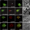
Dual-Localization Images of GFP-eIF4A-III with Several Nuclear Marker Proteins. (A) Coilin-RFP (Cajal body). (B) Fibrillarin-RFP (nucleolus). (C) CypRS64-RFP (splicing speckles). (D) RNPS1-RFP (splicing speckles) in Col-0 cells. Single confocal sections of GFP and RFP channels, merged fluorescent images, and corresponding bright-field images shown. Arrows indicate nucleoli; arrowheads indicate Cajal body. Bar = 10 μm.
Figure 4.
Figure 4.
Co-localisation of elF4A-III with Y14 and Mago proteins. Dual-localization images of eIF4A-III-RFP with GFP-Y14 (A) and GFP-Mago (B) in Col-0 cells. Single confocal sections of GFP and RFP channels and merged fluorescent images are shown. Bar = 4 μm.
Figure 5.
Figure 5.
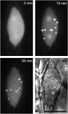
Dynamics of Speckle Formation in Nuclei of Col-0 Culture Cells Transiently Expressing GFP-eIF4A-III Placed on a Microscope Glass Slide with a Cover Slip. Two nucleoli and many bright splicing speckles can be seen at 15 min. The bottom right panel shows a bright-field image of the same nucleus. Bar = 10 μm.
Figure 6.
Figure 6.
Reversible effect of hypoxia treatment on elF4A-III localisation. Reversible dynamics of eIF4A-III in transiently expressing Col-0 culture cells placed in a modified gas-permeable chamber with a gate to block gas exchange. Note change from nucleoplasmic location in three nuclei ([A], 0 min) to speckles under hypoxia stress ([B], 60 min) and following recovery to nucleoplasmic pattern after 30 min in aerated conditions ([C], 90 min). Bar = 10 μm.
Figure 7.
Figure 7.
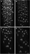
Hypoxia caused transition from diffuse nucleoplasmic labelling to accumulation in speckles in nuclei of root cells. Dynamics of eIF4A-III in Arabidopsis roots placed in a gas-permeable chamber: (A) and (C) with adequate aeration, and (B) and (D) in hypoxic conditions with 60 min of incubation. (A) and (B) show an area close to the root tip, and (C) and (D) show the root elongation zone of the same root. Nucleoplasmic pattern with less signal in the nucleoli can be seen in (A) and (B); some nuclei in (A) have very intense fluorescence. Faint nucleoplasm with bright speckles can be seen in nuclei in (B) and (D). Bar = 10 μm.
Figure 8.
Figure 8.
The Effect of Inhibition of Respiration by Azide on Localization of eIF4A-III in Hypocotyl Cells Expressing GFP-eIF4A-III. Control cells (A), 1 mM azide (B), and 100 mM azide (C) treatment for 30 min. Nucleoplasmic labeling in control cells (A) changed to accumulation in nucleoli and speckles at low concentration (1 mM) of sodium azide (B), while a higher concentration of the azide (100 mM) caused predominant accumulation of the protein in the nucleoli (C). Bar = 100 μm.
Figure 9.
Figure 9.
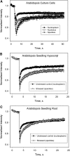
Intranuclear Mobility of Different Pools of GFP-eIF4A-III and FRAP Analysis of Different Nuclear Fractions. The background fluorescence was recorded, followed by photobleaching to significantly reduce the fluorescence intensity, and the fluorescence recovery was recorded as unbleached molecules diffused into the bleached area. Each line plotted using mean values ± se of 10 individual experiments. Formal kinetic parameters of recovery (maximal recovery, rate constant, and recovery half-time) were estimated using exponential model curves based on equation of one-phase exponential formation/association. (A) Diffuse nucleoplasmic, nucleolar, and speckles fractions in culture cells transiently expressing GFP-eIF4A-III. (B) Nucleoplasmic and speckles fractions of GFP-eIF4A-III in control and stressed hypocotyl cells of stably transformed Arabidopsis plants. (C) Nucleoplasmic and speckles fractions of GFP-eIF4A-III in control and stressed root cells of stably transformed Arabidopsis plants.

Comment in

References

    1. Alastalo, T.P., Hellesuo, M., Sandqvist, A., Hietakangas, V., Kallio, M., and Sistonen, L. (2003). Formation of nuclear stress granules involves HSF2 and coincides with the nucleolar localization of Hsp70. J. Cell Sci. 116 3557–3570. - PubMed
    1. Ali, G.S., and Reddy, A.S. (2006). ATP, phosphorylation and transcription regulate the mobility of plant splicing factors. J. Cell Sci. 119 3527–3538. - PubMed
    1. Andersen, C.B., Ballut, L., Johansen, J.S., Chamieh, H., Nielsen, K.H., Oliveira, C.L., Pedersen, J.S., Seraphin, B., Le Hir, H., and Andersen, G.R. (2006). Structure of the exon junction core complex with a trapped DEAD-box ATPase bound to RNA. Science 313 1968–1972. - PubMed
    1. Ares, M., Jr., and Proudfoot, N.J. (2005). The spanish connection: transcription and mRNA processing get even closer. Cell 120 163–166. - PubMed
    1. Bailey-Serres, J., and Chang, R. (2005). Sensing and signalling in response to oxygen deprivation in plants and other organisms. Ann. Bot. (Lond.) 96 507–518. - PMC - PubMed

Publication types

MeSH terms