

Valves of the deep venous system: an overlooked risk factor
- PMID: 19436051
- PMCID: PMC2723019
- DOI: 10.1182/blood-2009-03-209981
Valves of the deep venous system: an overlooked risk factor
Abstract
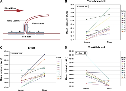
Deep venous valves are frequent sites of deep venous thrombosis initiation. However, the possible contribution of the valvular sinus endothelium has received little attention in studies of thrombosis risk. We hypothesized that the endothelium of valve sinus differs from that of vein lumen with up-regulation of anticoagulant and down-regulation of procoagulant activities in response to the local environment. In pursuit of this hypothesis, we quantified endothelial protein C receptor (EPCR), thrombomodulin (TM), and von Willebrand factor (VWF) by immunofluorescence in great saphenous veins harvested at cardiac bypass surgery. We found significantly increased expression of EPCR and TM in the valvular sinus endothelium as opposed to the vein lumenal endothelium, and the opposite pattern with VWF (paired t test for TM and EPCR, each P < .001; for VWF, P = .01). These data support our hypothesis and suggest that variation in valvular sinus thromboresistance may be an important factor in venous thrombogenesis.
Figures
Comment in
-
Is Virchow's triad complete?Blood. 2009 Aug 6;114(6):1138-9. doi: 10.1182/blood-2009-05-223511. Blood. 2009. PMID: 19661277 No abstract available.
Similar articles
-
The endothelial microenvironment in the venous valvular sinus: thromboresistance trends and inter-individual variation.Histochem Cell Biol. 2011 Feb;135(2):141-52. doi: 10.1007/s00418-011-0783-5. Epub 2011 Feb 6. Histochem Cell Biol. 2011. PMID: 21298440 Free PMC article.
-
Free fatty acids inhibit TM-EPCR expression through JNK pathway: an implication for the development of the prothrombotic state in metabolic syndrome.J Thromb Thrombolysis. 2012 Nov;34(4):468-74. doi: 10.1007/s11239-012-0793-8. J Thromb Thrombolysis. 2012. PMID: 22903729
-
Arterial flow conditions downregulate thrombomodulin on saphenous vein endothelium.Circulation. 1999 Mar 2;99(8):1047-53. doi: 10.1161/01.cir.99.8.1047. Circulation. 1999. PMID: 10051299
-
Venous valve hypoxia as a possible mechanism of deep vein thrombosis: a scoping review.Int Angiol. 2024 Jun;43(3):309-322. doi: 10.23736/S0392-9590.24.05170-8. Epub 2024 Jun 12. Int Angiol. 2024. PMID: 38864688
-
To what extent might deep venous thrombosis and chronic venous insufficiency share a common etiology?Int Angiol. 2009 Aug;28(4):254-68. Int Angiol. 2009. PMID: 19648868 Review.
Cited by
-
Deep vein thrombosis: a clinical review.J Blood Med. 2011;2:59-69. doi: 10.2147/JBM.S19009. Epub 2011 Apr 29. J Blood Med. 2011. PMID: 22287864 Free PMC article.
-
Tumor-derived tissue factor-positive microparticles and venous thrombosis in cancer patients.Blood. 2013 Sep 12;122(11):1873-80. doi: 10.1182/blood-2013-04-460139. Epub 2013 Jun 24. Blood. 2013. PMID: 23798713 Free PMC article. Review.
-
In silico analyses of blood flow and oxygen transport in human micro-veins and valves.Clin Hemorheol Microcirc. 2022;81(1):81-96. doi: 10.3233/CH-211345. Clin Hemorheol Microcirc. 2022. PMID: 35034895 Free PMC article.
-
Role of tissue factor in venous thrombosis.Annu Rev Physiol. 2011;73:515-25. doi: 10.1146/annurev-physiol-042210-121137. Annu Rev Physiol. 2011. PMID: 20690821 Free PMC article. Review.
-
Venous Thromboembolism: Review of Clinical Challenges, Biology, Assessment, Treatment, and Modeling.Ann Biomed Eng. 2024 Mar;52(3):467-486. doi: 10.1007/s10439-023-03390-z. Epub 2023 Nov 1. Ann Biomed Eng. 2024. PMID: 37914979 Review.
References
-
- Paterson JC, McLachlin J. Precipitating factors in venous thrombosis. Surg Gynecol Obstet. 1954;98:96–102. - PubMed
-
- Gottlob M, May R. Part III: pathologic venous valves. In: Gottlob R, May R, editors. Venous Valves: Morphology, Function, Radiology, Surgery. New York: Springer-Verlag; 1986. pp. 82–92.
-
- Lund FL, Diener L, Ericsson JLE. Postmortem intraosseous phlebography as an aid in studies of venous thromboembolism: with application on a geriatric clientele. Angiology. 1969;20:155–176. - PubMed
Publication types
MeSH terms
Substances
Grants and funding
LinkOut - more resources
Full Text Sources
Other Literature Sources
Medical
Miscellaneous
