. 2009 Jun;119(6):1626-37.
doi: 10.1172/JCI35288. Epub 2009 May 11.

Liver X receptors contribute to the protective immune response against Mycobacterium tuberculosis in mice

Affiliations

Liver X receptors contribute to the protective immune response against Mycobacterium tuberculosis in mice

Hannelie Korf et al. J Clin Invest. 2009 Jun.

Abstract

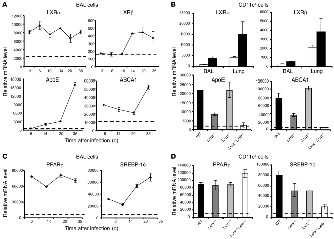
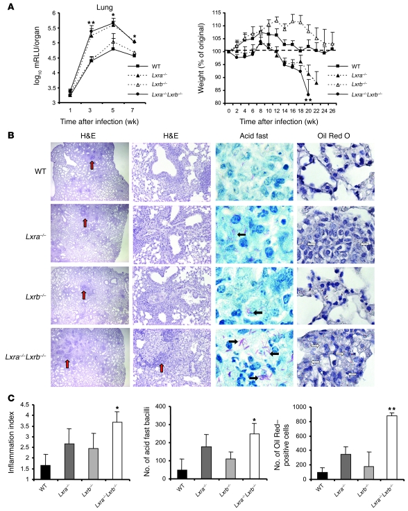
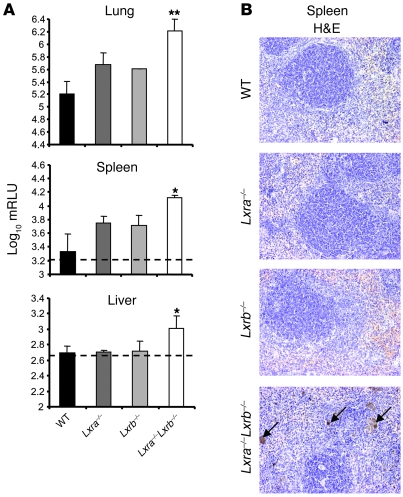
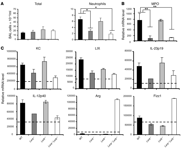
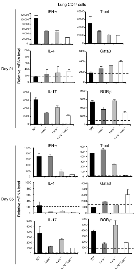
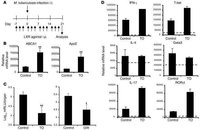
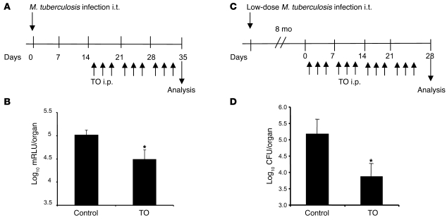
Liver X receptors (LXRs) are key regulators of macrophage function, controlling transcriptional programs involved in lipid homeostasis and inflammation. However, exactly how LXRs modulate inflammation during infection remains unknown. To explore this, we used a mouse model of Mycobacterium tuberculosis infection. Upon intratracheal infection with M. tuberculosis, LXRs and LXR target genes were induced in CD11c+ lung and alveolar cells. Furthermore, mice deficient in both LXR isoforms, LXRalpha and LXRbeta (Lxra-/-Lxrb-/- mice), were more susceptible to infection, developing higher bacterial burdens and an increase in the size and number of granulomatous lesions. Interestingly, mice solely deficient in LXRalpha, but not those lacking only LXRbeta, mirrored the susceptibility of the Lxra-/-Lxrb-/- animals. Lxra-/-Lxrb-/- mice failed to mount an effective early neutrophilic airway response to infection and showed dysregulation of both pro- and antiinflammatory factors in CD11c+ lung cells. T cell responses were strongly affected in Lxra-/-Lxrb-/- mice, showing near-complete abrogation of the infection-induced Th1 function - and even more so Th17 function - in the lungs. Treatment of WT mice with the LXR agonists TO901317 and GW3965 resulted in a 10-fold decrease of the pulmonary bacterial burden and a comparable increase of Th1/Th17 function in the lungs. The dependence of LXR signaling on the neutrophil IL-17 axis represents what we believe to be a novel function for these nuclear receptors in resistance to M. tuberculosis infection and may provide a new target for therapeutics.


Figures

Figure 1
Figure 1. Characteristic features of the murine pulmonary tuberculosis model.
(A) Bacterial counts in the lungs and spleen at different time points after i.t. instillation of luminescent M. tuberculosis (1 × 10⁴ CFU). Values are expressed as log10 mRLU per organ (n = 4). Dotted lines indicate the detection limit of the bioluminescence assay. (B) RT-qPCR analysis of the indicated inflammatory mediators within BAL cells. Data are expressed as relative mRNA levels, normalized against reference the housekeeping gene. Mice were tested individually (n = 4). Dotted lines denote mRNA levels obtained from background-matched naive BAL cells. (C) Cellular infiltration in the airways after exposure to the pathogen. Shown are absolute numbers of total cells, mononuclear cells, neutrophils, and lymphocytes. Dotted lines denote cell numbers in naive animals. Data in C are mean ± SEM (n = 5).
Figure 2
Figure 2. Expression of LXRs and LXR-dependent genes during M. tuberculosis infection.
(A and C) RT-qPCR analysis of expression levels of LXRα and LXRβ isoforms, LXR-dependent target genes, and PPARγ and SREBP-1c within BAL cells isolated at the indicated times after infection. Dotted lines denote mRNA levels obtained from background-matched naive BAL cells. Mice were tested individually (n = 4–5). (B and D) After 3 weeks of infection, CD11c+ cells were isolated from the BAL, and lung tissue digests and relative mRNA levels of the indicated parameters were determined by RT-qPCR. (B, top) Infected (black bars) CD11c+ BAL and lung cells or naive controls (white bars) from WT mice. (B, bottom, and D) Transcriptional analysis of CD11c+ BAL cells isolated from infected WT, Lxra–/–, Lxrb–/–, and Lxra–/–Lxrb–/– mice (n = 5). Dotted lines denote mRNA levels obtained from background-matched naive CD11c+ BAL cells.
Figure 3
Figure 3. Mice lacking LXRs are more susceptible to M. tuberculosis infection.
(A) Changes in bacterial load in the lungs and total body mass of WT, Lxra–/–, Lxrb–/–, and Lxra–/–Lxrb–/– mice infected i.t. with M. tuberculosis (1 × 10⁴ CFU). Values are expressed as log10 mRLU per organ and as percent of original weight (n = 5–7). *P < 0.05, **P < 0.01, Lxra–/–Lxrb–/– versus WT. (B) Histological analysis of H&E-stained lungs sections at 5 weeks after infection revealed more extensive granulomatous inflammation (red arrows) as well as total inflammation in the in Lxra–/–Lxrb–/– mice. Acid fast staining of the sections showed the presence of mycobacteria as pink rods. Frequent events of multiple bacteria per cell were observed in the lung sections from Lxra–/– and Lxra–/–Lxrb–/– mice (black arrows). Foamy macrophages containing multiple red lipid droplets after staining with Oil Red O was most prominent in Lxra–/– and Lxra–/–Lxrb–/– mice (white arrows). Original magnification (left to right), ×20, ×100, ×1,000, ×600. (C) Quantification of the lung inflammation index, number of bacilli, and Oil Red–positive cells, as described in Methods (n = 5). *P < 0.05, **P < 0.01 versus WT.
Figure 4
Figure 4. Infection of Lxra–/–Lxrb–/– mice results in disseminated systemic infection.
(A) Bacterial load in the lungs, spleens, and livers of WT, Lxra–/–, Lxrb–/–, and Lxra–/–Lxrb–/– mice infected i.t. with luminescent M. tuberculosis (1 × 10⁵ CFU) for 5 weeks. Values are expressed as log10 mRLU per organ (n = 3). Dotted lines indicate the detection limit of the bioluminescence assay. *P < 0.05, **P < 0.01 versus WT. (B) Histological analysis of spleen sections prepared after 5 weeks of infection revealed the presence in Lxra–/–Lxrb–/– mice of multiple macrophages with a brown reaction product after standard H&E staining (arrows). Original magnification, ×100.
Figure 5
Figure 5. LXR-dependent regulation of the innate immune response after i.t. challenge with M. tuberculosis.
WT, Lxra–/–, Lxrb–/–, and Lxra–/–Lxrb–/– mice were infected i.t. with luminescent M. tuberculosis (1 × 10⁴ CFU) and sacrificed 7 days after infection. (A) Differential cell infiltration in the BAL. Shown are the absolute numbers of total cells and neutrophils. Data in A are mean ± SEM (n = 5). (B) Relative mRNA levels of MPO from total lung tissue samples, as determined by RT-qPCR (n = 5). (C) CD11c+ cells were isolated 7 days after infection from lung tissue digests, and relative mRNA levels were determined by RT-qPCR. Dotted lines denote values obtained from CD11c+ cells of naive mice. Data are expressed as relative mRNA levels, normalized against reference housekeeping genes (n = 5), and are representative of 2 separate experiments. Arg, arginase. *P < 0.05, **P < 0.01.
Figure 6
Figure 6. The role of LXRs in the modulation of T cell function during M. tuberculosis infection.
WT, Lxra–/–, Lxrb–/–, and Lxra–/–Lxrb–/– mice were infected i.t. with luminescent M. tuberculosis (1 × 10⁴ CFU). After 21 and 35 days of infection, CD4+ lung T cells were isolated, and the relative mRNA levels of Th1 (IFN-γ and T-bet), Th2 (IL-4 and Gata3), and Th17 (IL-17 and RORγt) markers were analyzed by RT-qPCR. Data are expressed as relative mRNA levels, normalized against reference housekeeping genes (n = 5). Results are representative of 2 separate experiments.
Figure 7
Figure 7. Prophylactic LXR agonist treatment protects mice against airway challenge with M. tuberculosis.
(A) Schedule of prophylactic treatment with LXR agonist TO91317 (TO) or GW3965 (GW) in conjunction with the i.t. M. tuberculosis challenge model. WT C57BL/6 mice were injected i.p. 3 times per week with 50 μg agonist. (B) RT-qPCR analysis of LXR-dependent target gene mRNA in total BAL cells from mock-infected animals using the same treatment schedule. (C) Bacterial load in the lungs of treated and control groups at day 21 after infection. No difference was observed in the vehicle-treated control group and the placebo-treated group (not shown). Values are expressed as log10 mRLU per organ (n = 5). (D) CD4+ lung T cells were isolated after 21 days of infection, and the relative mRNA levels of Th1, Th2, and Th17 markers (described in Figure 6) were analyzed by RT-qPCR. Data are expressed as relative mRNA levels, normalized against reference housekeeping genes (n = 5). Results are representative of 2 separate experiments. *P < 0.05, **P < 0.01 versus control.
Figure 8
Figure 8. Therapeutic treatment with LXR agonist protects mice against airway challenge with M. tuberculosis.
(A and C) Schedule of therapeutic treatment with TO91317 in conjunction with the i.t. M. tuberculosis challenge model. WT C57BL/6 mice were injected i.p. 3 times per week with 50 μg TO91317. (B) Bacterial load in the lungs at 35 days after infection. Values are expressed as log10 mRLU per organ (n = 5). (D) Bacterial load in the lungs at 28 days after infection. Values are expressed as log10 CFU per organ (n = 5). *P < 0.05, **P < 0.01 versus control.

References

    1. WHO. 2005. Tuberculosis [fact sheet]. Revised March 2007. http://www.who.int/mediacentre/factsheets/fs104/en.
    1. Manabe Y.C., Bishai W.R. Latent Mycobacterium tuberculosis-persistence, patience, and winning by waiting. Nat. Med. 2000;6:1327–1329. doi: 10.1038/82139. - DOI - PubMed
    1. Rengarajan J., Bloom B.R., Rubin E.J. Genome-wide requirements for Mycobacterium tuberculosis adaptation and survival in macrophages. Proc. Natl. Acad. Sci. U. S. A. 2005;102:8327–8332. doi: 10.1073/pnas.0503272102. - DOI - PMC - PubMed
    1. Schnappinger D., et al. Transcriptional adaptation of Mycobacterium tuberculosis within macrophages: insights into the phagosomal environment. . J. Exp. Med. 2003;198:693–704. doi: 10.1084/jem.20030846. - DOI - PMC - PubMed
    1. Voskuil M.I., et al. Inhibition of respiration by nitric oxide induces a Mycobacterium tuberculosis dormancy program. J. Exp. Med. 2003;198:705–713. doi: 10.1084/jem.20030205. - DOI - PMC - PubMed

Publication types

MeSH terms