. 2009 Jun;119(6):1546-57.
doi: 10.1172/JCI35722. Epub 2009 May 11.

Prevention of hepatocarcinogenesis and increased susceptibility to acetaminophen-induced liver failure in transaldolase-deficient mice by N-acetylcysteine

Affiliations

Prevention of hepatocarcinogenesis and increased susceptibility to acetaminophen-induced liver failure in transaldolase-deficient mice by N-acetylcysteine

Robert Hanczko et al. J Clin Invest. 2009 Jun.

Abstract

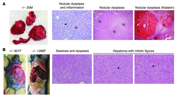
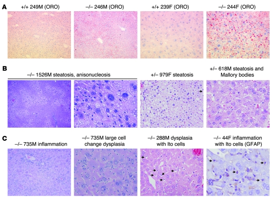
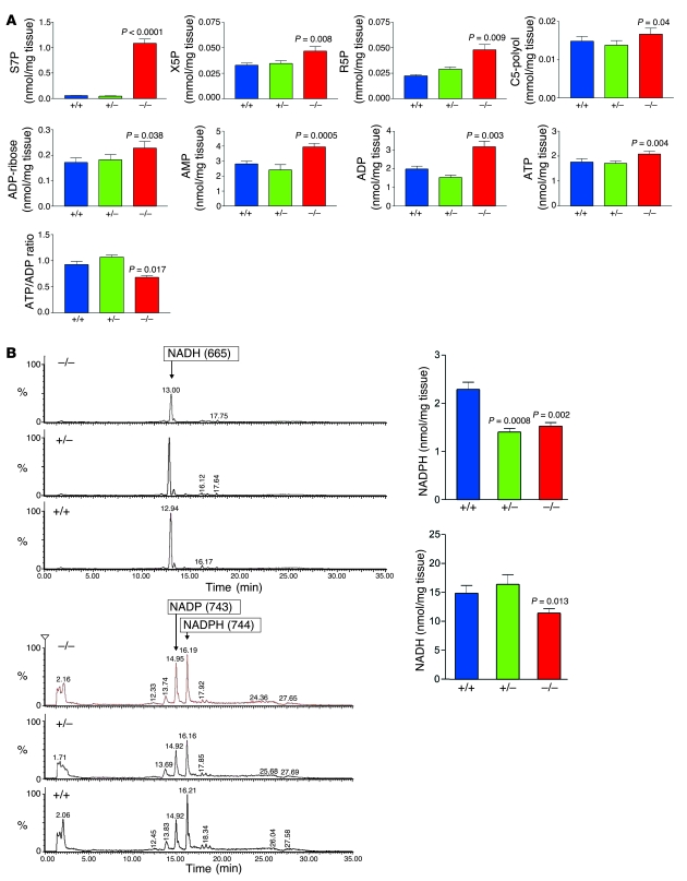
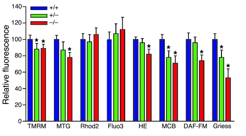
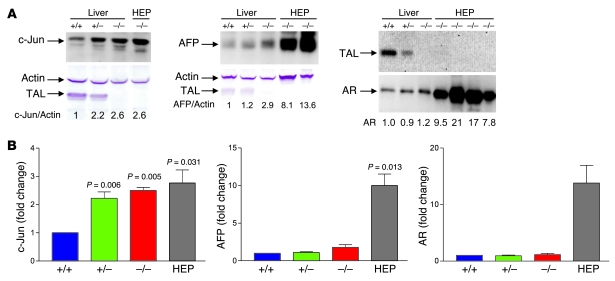
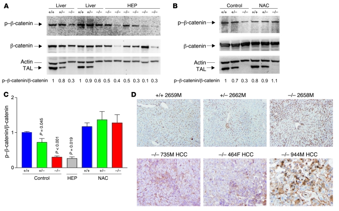
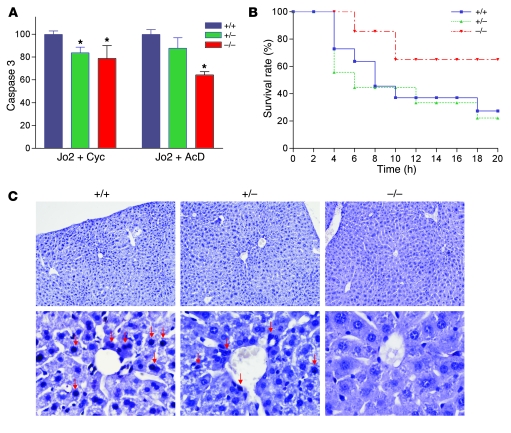
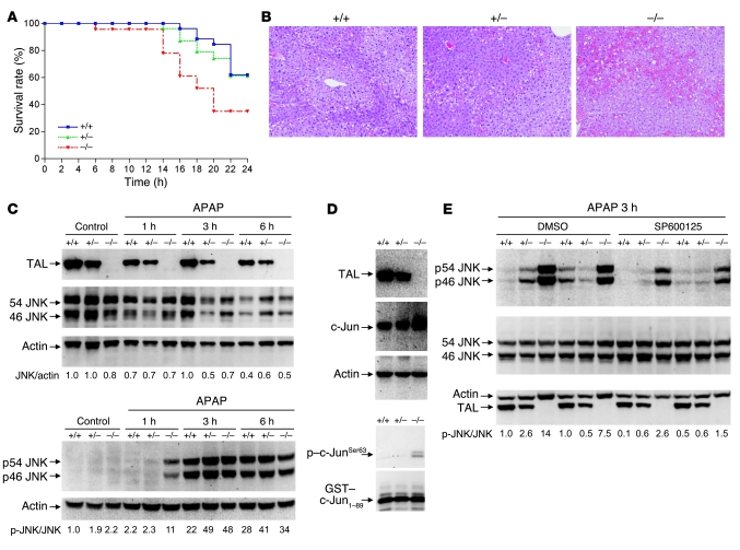
Although oxidative stress has been implicated in acute acetaminophen-induced liver failure and in chronic liver cirrhosis and hepatocellular carcinoma (HCC), no common underlying metabolic pathway has been identified. Recent case reports suggest a link between the pentose phosphate pathway (PPP) enzyme transaldolase (TAL; encoded by TALDO1) and liver failure in children. Here, we show that Taldo1-/- and Taldo1+/- mice spontaneously developed HCC, and Taldo1-/- mice had increased susceptibility to acetaminophen-induced liver failure. Oxidative stress in Taldo1-/- livers was characterized by the accumulation of sedoheptulose 7-phosphate, failure to recycle ribose 5-phosphate for the oxidative PPP, depleted NADPH and glutathione levels, and increased production of lipid hydroperoxides. Furthermore, we found evidence of hepatic mitochondrial dysfunction, as indicated by loss of transmembrane potential, diminished mitochondrial mass, and reduced ATP/ADP ratio. Reduced beta-catenin phosphorylation and enhanced c-Jun expression in Taldo1-/- livers reflected adaptation to oxidative stress. Taldo1-/- hepatocytes were resistant to CD95/Fas-mediated apoptosis in vitro and in vivo. Remarkably, lifelong administration of the potent antioxidant N-acetylcysteine (NAC) prevented acetaminophen-induced liver failure, restored Fas-dependent hepatocyte apoptosis, and blocked hepatocarcinogenesis in Taldo1-/- mice. These data reveal a protective role for the TAL-mediated branch of the PPP against hepatocarcinogenesis and identify NAC as a promising treatment for liver disease in TAL deficiency.


Figures

Figure 1
Figure 1. Spontaneous development of dysplastic nodules, cirrhosis, and HCC in Taldo1–/– mice.
(A) Dysplastic nodules and cirrhosis in Taldo1–/– (–/–) mice. Macroscopic and microscopic images of 52-week-old Taldo1–/– mouse 20M stained with H&E and Klatskin trichrome stain. Arrow denotes inflammatory cells surrounding nodules (N). Klatskin trichrome staining of the liver showed interstitial deposition of collagen (blue). Hepatocytes within the nodules were avidly stained red. Original magnification, left to right: ×100, ×40, ×40. (B) Macroscopic images of HCC in a 74-week-old (931F) and an 82 week-old (1266F) Taldo1–/– mice, and microscopic images of steatosis, dysplasia, mitotic figures (arrows), and hepatoma in 74-week-old Taldo1–/– mouse 931F. Original magnification, left to right: ×100, ×100, ×200.
Figure 2
Figure 2. NAFLD and NASH with dysplasia in 10- to 12-week-old Taldo1–/– mice.
(A) Detection of lipid droplets with oil-red-O (ORO) staining in frozen liver sections of Taldo1–/– and Taldo1+/+ (+/+) littermates. Original magnification, left to right: ×100, ×100, ×400, ×400. (B) Centrolobular, zonal, or diffuse steatosis and Mallory bodies in Taldo1–/– and Taldo1+/– (+/–) mice. Original magnification, left to right: ×40, ×400, ×400, ×400. (C) Steatosis, inflammation, liver cell dysplasia with large cell change, and expansion of fat-storing hepatic stellate or Ito cells (arrows). Ito cells were also identified by expression of glial fibrillary acidic protein (GFAP). Original magnification, left to right: ×100, ×400, ×400, ×600.
Figure 3
Figure 3. LC-MS/MS analysis of PPP sugars and nucleotides in the liver.
(A) Cumulative analysis of S7P, X5P, R5P, C5-polyol, ADP-ribose, AMP, ADP, and ATP levels as well as the ATP/ADP ratio in the livers of 14 sets (6 male, 8 female) of Taldo1–/–, Taldo1+/+, and Taldo1+/– littermates. All samples were analyzed after methanol/water extraction by LC-MS/MS (25). Sugar phosphates and nucleotides were detected in the same sample. (B) Quantitative recovery of NADH and NADPH was achieved as described in Methods. This method allowed parallel detection of NADH, NADP, and NADPH, as shown by representative MS/MS chromatograms in liver samples from female littermates. Cumulative analysis of NADPH and NADH levels in 12 female littermates 5–6 weeks of age is shown at right. P values compared with Taldo1+/+ mice are shown.
Figure 4
Figure 4. Flow cytometry analysis of mitochondrial function and NO production by isolated hepatocytes.
Mouse hepatocytes were isolated from 8- to 10-week-old Taldo1+/+, Taldo1+/–, and Taldo1–/– female littermates by a 2-step perfusion procedure as described in Methods. Δψm by TMRM, mitochondrial mass by MitoTracker Green (MTG), mitochondrial and cytoplasmic Ca2+ levels by Rhod-2 and Fluo-3, ROI production by H&E, and NO production by DAF-FM fluorescence were assessed in annexin V–negative cells (72). GSH levels were assessed by monochlorobimane (MCB) fluorescence. NO levels were also assessed by the Griess reaction. Data are mean of 4 or more independent experiments normalized to Taldo1+/+ hepatocyte values, set as 100%. *P < 0.05 versus Taldo1+/+.
Figure 5
Figure 5. Expression of c-Jun, AFP, and AR in Taldo1+/+, Taldo1+/–, and Taldo1–/– liver tissues from 5-week-old female littermates and in hepatomas (HEP) from Taldo1–/– mice.
While c-Jun and AFP were detected in whole cell lysates, AR was analyzed in cytosolic extracts (73). (A) Western blot detection of c-Jun, AFP, and AR expression relative to β-actin in livers and hepatoma tissues. Numbers below blots indicate densitometry values, which were normalized to those of Taldo1+/+ livers, set at 1.0. (B) Cumulative analyses of c-Jun/actin, AFP/actin, and AR/actin levels by Western blot in Taldo1–/– hepatomas and tumor-free Taldo1–/– and Taldo1+/– livers relative to Taldo1+/+ liver. Data represent mean ± SEM of 4 littermate sets and 4 hepatomas. P values denoting significant differences versus Taldo1+/+ mice are shown.
Figure 6
Figure 6. Effect of TAL deficiency on phosphorylation and intracellular localization of β-catenin in Taldo1+/+, Taldo1+/–, and Taldo1–/– liver tissues from 10- to 12-week-old littermates and hepatomas from Taldo1–/– mice.
(A) Western blot detection of phosphorylated β-catenin (p–β-catenin) relative to β-catenin in liver and hepatoma tissues. As a loading control, p–β-catenin and β-catenin levels were normalized to actin for each sample. (B) Effect of lifelong NAC treatment on p–β-catenin/β-catenin levels in liver tissues of 12-week-old littermates. Numbers below blots show p–β-catenin/β-catenin ratios (i.e., [p–β-catenin/actin]/[β-catenin/actin]), which were normalized to those of Taldo1+/+ livers, set at 1.0. (C) Cumulative analyses of p–β-catenin/β-catenin levels in liver tissues of 12-week-old littermates, hepatomas, and liver tissues from NAC-treated 10- to 12-week-old littermates. Data represent mean ± SEM of 4 littermate sets and 13 hepatomas. P values denoting significant differences versus control-treated Taldo1+/+ mice are shown. (D) Immunohistochemical analysis of β-catenin expression in liver tissues of 12-week-old littermates and in Taldo1–/– livers with HCC. Original magnification, ×100.
Figure 7
Figure 7. Resistance of Taldo1–/– hepatocytes to Fas-induced apoptosis.
(A) Fas-induced apoptosis of hepatocytes in vitro by activation of caspase 3. Hepatocytes were incubated as described in Methods with anti-Fas antibody (Jo2, 0.5 μg/ml) and 50 μg/ml cycloheximide (Cyc) or 50 ng/ml actinomycin D (AcD). Mean ± SEM values from 4 litters per independent experiment are expressed relative to hepatocytes from Taldo1+/+ littermates, set as 100%. *P < 0.05 versus Taldo1+/+. (B) Survival of 8- to 10-week-old Taldo1+/+ (n = 22), Taldo1+/– (n = 15), and Taldo1–/– littermate mice (n = 14) following i.p. injection with Jo2 antibody (10 μg/30 g body weight). Log-rank test showed increased survival of Taldo1–/– mice compared with Taldo1+/+ mice (P < 0.0001). No significant difference was observed between the Taldo1+/– and Taldo1+/+ groups. (C) TUNEL staining of liver sections 4 hours after i.p. injection with Jo2 antibody. Condensed and fragmented apoptotic nuclei (red arrows) in Taldo1+/+ and Taldo1+/– livers were visualized by staining with Vector Black substrate. Sections were counterstained with hematoxylin. Original magnification, ×100 (top row); ×400 (bottom row).
Figure 8
Figure 8. Increased susceptibility of Taldo1–/– mice to liver failure induced by APAP.
(A) Survival of Taldo1+/+ (n = 26), Taldo1+/– (n = 28), and Taldo1–/– littermates (n = 23) injected with 800 mg/kg APAP. Log-rank test showed reduced survival of Taldo1–/– compared with Taldo1+/+ mice (P = 0.027). No significant difference was observed between the Taldo1+/– and Taldo1+/+ groups. (B) H&E-stained liver sections obtained 6 hours after APAP injection. Hemorrhagic necrosis, characterized by hepatocyte vacuolization and extravasation of erythrocytes, was enhanced in Taldo1–/– liver. Original magnification, ×100. (C) Western blot detection of 46- and 54-kDa JNK and their state of phosphorylation (p46 and p54 JNK) in APAP-injected and untreated control mice. Numbers below blots show p-JNK/JNK levels, which were determined relative to actin and normalized to untreated Taldo1+/+ protein lysates, set as 1.0. (D) Assessment of JNK activity by in vitro phosphorylation of GST–c-Jun1–89 fusion protein in APAP-treated liver. TAL, c-Jun, and actin levels were detected by Western blot of liver cell lysates. In vitro phosphorylation of GST–c-Jun was detected by Western blot analysis using anti–phospho–c-JunSer63 antibody. (E) Effect of SP600125 on APAP-induced activation of JNK. Littermate 12-week-old mice were pretreated with SP600125 or DMSO control as described in Methods 1 hour prior to APAP exposure. Numbers below blots show p-JNK/JNK levels 3 hours after APAP treatment; values were determined relative to actin and normalized to untreated Taldo1+/+ protein lysates, set as 1.0.

Comment in

Similar articles

Cited by

References

    1. Ahn J., Flamm S.L. Hepatocellular carcinoma. Dis. Mon. 2004;50:556–573. doi: 10.1016/j.disamonth.2004.10.001. - DOI - PubMed
    1. Stickel F., Schuppan D., Hahn E.G., Seitz H.K. Cocarcinogenic effects of alcohol in hepatocarcinogenesis. Gut. 2002;51:132–139. doi: 10.1136/gut.51.1.132. - DOI - PMC - PubMed
    1. El Serag H.B. Hepatocellular carcinoma and hepatitis C in the United States. Hepatology. 2002;36:S74–S83. doi: 10.1002/hep.1840360710. - DOI - PubMed
    1. Nguyen M.H., Keeffe E.B. Screening for hepatocellular carcinoma. J. Clin. Gastroenterol. 2002;35:S86–S91. doi: 10.1097/00004836-200211002-00004. - DOI - PubMed
    1. Moradpour D., Blum H.E. Pathogenesis of hepatocellular carcinoma. Eur. J. Gastroenterol. Hepatol. 2005;17:477–483. doi: 10.1097/00042737-200505000-00002. - DOI - PubMed

Publication types

MeSH terms