. 2009 Jun;119(6):1688-95.
doi: 10.1172/JCI36891. Epub 2009 May 11.

Capsid antigen presentation flags human hepatocytes for destruction after transduction by adeno-associated viral vectors

Affiliations

Capsid antigen presentation flags human hepatocytes for destruction after transduction by adeno-associated viral vectors

Gary C Pien et al. J Clin Invest. 2009 Jun.

Abstract

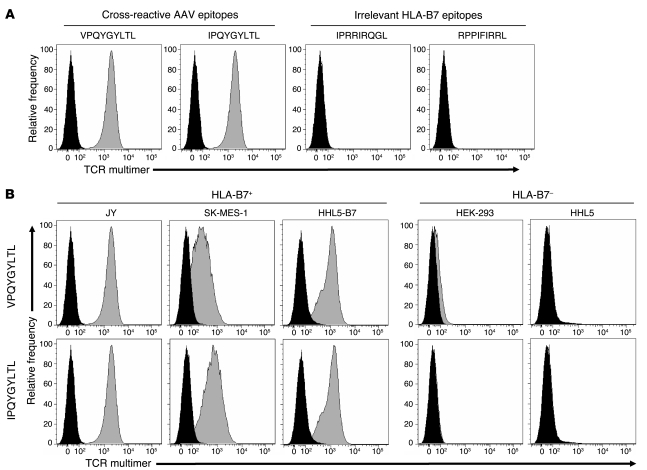
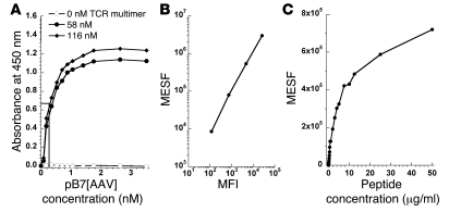
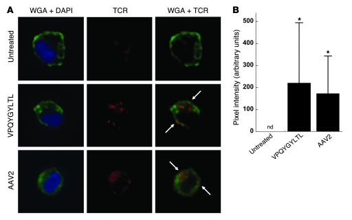
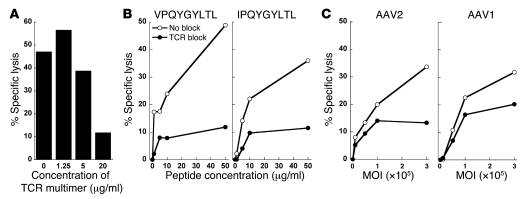
Adeno-associated virus (AAV) vectors are effective gene delivery vehicles mediating long-lasting transgene expression. Data from a clinical trial of AAV2-mediated hepatic transfer of the Factor IX gene (F9) into hemophilia B subjects suggests that CTL responses against AAV capsid can eliminate transduced hepatocytes and prevent long-term F9 expression. However, the capacity of hepatocytes to present AAV capsid-derived antigens has not been formally demonstrated, nor whether transduction by AAV sensitizes hepatocytes for CTL-mediated destruction. To investigate the fate of capsids after transduction, we engineered a soluble TCR for the detection of capsid-derived peptide:MHC I (pMHC) complexes. TCR multimers exhibited antigen and HLA specificity and possessed high binding affinity for cognate pMHC complexes. With this reagent, capsid pMHC complexes were detectable by confocal microscopy following AAV-mediated transduction of human hepatocytes. Although antigen presentation was modest, it was sufficient to flag transduced cells for CTL-mediated lysis in an in vitro killing assay. Destruction of hepatocytes was inhibited by soluble TCR, demonstrating a possible application for this reagent in blocking undesirable CTL responses. Together, these studies provide a mechanism for the loss of transgene expression and transient elevations in aminotransferases following AAV-mediated hepatic gene transfer in humans and a potential therapeutic intervention to abrogate these limitations imposed by the host T cell response.


Figures

Figure 1
Figure 1. Antigen and HLA specificities of TCR multimer staining.
(A) To demonstrate antigen specificity, HLA-B*0702+ JY cells were pulsed with 5 μg/ml of either cross-reactive capsid epitopes derived from AAV2 (VPQYGYLTL) or AAV8 (IPQYGYLTL) or irrelevant HLA-B*0702–restricted epitopes derived from HIV (IPRRIRQGL) or EBV (RPPIFIRRL). After 2 hours of incubation, cells were stained with TCR multimer for flow cytometry. Histograms depict unpulsed cells (black) and peptide-pulsed cells (gray). (B) To demonstrate HLA specificity, either HLA-matched (JY, SK-MES-1, and HHL5-B7) or HLA-mismatched (HEK-293 and HHL5) cells were pulsed with 5 μg/ml of indicated AAV capsid peptide, then stained with TCR multimer. Histograms depict unpulsed cells (black) and peptide-pulsed cells (gray).
Figure 2
Figure 2. Estimation of TCR multimer binding affinity and limit of detection.
(A) TCR multimer was used to coat microtiter wells at 0 nM (open circles), 58 nM (filled circles), or 116 nM (filled diamonds). Biotinylated pB7[AAV] was then added at the indicated concentrations, followed by streptavidin-conjugated HRP. Absorbance at 450 nm was measured following addition of o-phenylenediamine substrate. Binding affinity (Kd) was estimated from the concentration of pB7[AAV] at which half-maximal detection occurred (EC50). (B) A set of APC calibration beads was used to set photomultiplier tube voltages on a FACS Canto II. Then, a calibration curve was generated using linear regression to calculate MESF from MFI (r2 ≥ 0.99). (C) After determining the MFI of peptide-pulsed HHL5-B7 hepatocytes stained with TCR multimer, the MESF was calculated and plotted against peptide concentration. The minimum number of pMHC complexes detectable by flow cytometry was estimated from the lowest MESF discernible from unpulsed cells.
Figure 3
Figure 3. Confocal microscopy of TCR multimer staining following AAV-mediated transduction.
(A) HHL5-B7 hepatocytes were cultured alone or in the presence of 10 μg/ml VPQYGYLTL capsid peptide or AAV2-F9 vector at 3 × 10⁵ MOI. Cells were then stained with WGA (green) to visualize the plasma membrane, DAPI (blue) to identify the nucleus, and TCR multimer (red) to stain pMHC complexes. Cells were visualized using spinning disk confocal microscopy, and representative cells are shown. Arrows indicate representative areas of TCR and plasma membrane colocalization (yellow). Total original magnification, ×63. (B) Staining intensity of colocalized signal from individual cells was quantitatively measured using Volocity and graphed as average intensity per cell ± SD (n = 17–25 cells per group). After normalization as detailed in Methods, the colocalized intensity was not detectable (nd) on untreated cells. *P < 0.004 versus untreated cells.
Figure 4
Figure 4. Cytotoxic activity against peptide-pulsed and AAV-transduced hepatocytes.
In separate experiments, the human hepatocyte cell line HHL5 expressing HLA-A*0101 was cultured with (A) 10 μg/ml of SADNNNSEY capsid peptide, or the HLA-B*0702–expressing HHL5-B7 cell line was cultured with (B) 3 × 10⁵ MOI AAV2-F9 vector or (C) 3 × 10⁵ MOI AAV2-empty capsids devoid of genomes. HLA-matched human PBMCs were added at the indicated effector/target (E:T) ratios in a CTL cytotoxicity assay. The percentage of specific lysis was calculated from the release of intracellular lactate dehydrogenase. To determine the effects of antigen load on CTL cytotoxicity, HHL5-B7 target cells were incubated with the indicated concentrations of VPQYGYLTL capsid peptide (D) or MOI of AAV2-F9 (E) overnight, then effectors were added at an effector/target ratio of 10:1. Effector cells were derived as follows: (AD) normal human donor PBMCs were independently expanded for 2 rounds with capsid peptide or AAV2-empty capsid and pooled for use as effector cells or (E) human PBMCs were expanded for 2 rounds with AAV2-empty capsid only.
Figure 5
Figure 5. TCR multimers block CTL cytotoxicity.
(A) Normal human donor PBMCs were expanded for 3 rounds with capsid peptide or AAV2-empty capsid and pooled for use as effector cells. HLA-matched HHL5-B7 target cells were transduced with AAV2-F9 at 3 × 10⁵ MOI. Effectors and target cells were then plated at an effector/target ratio of 10:1 in the absence or presence of indicated concentrations of TCR multimer. (B and C) In a separate experiment, HHL5-B7 hepatocytes were cultured with either AAV-derived capsid peptides (B) or AAV vectors (C) at the indicated concentrations. The following day, effector cells were added at an effector/target ratio of 10:1 in the absence (open circles) or presence (filled circles) of 20 μg/ml TCR multimer. Effectors were derived from pooled human PBMCs expanded for 1 round with capsid peptide or AAV2-empty capsid.

References

    1. Manno C.S., et al. Successful transduction of liver in hemophilia by AAV-Factor IX and limitations imposed by the host immune response. Nat. Med. 2006;12:342–347. doi: 10.1038/nm1358. - DOI - PubMed
    1. Mingozzi F., et al. CD8(+) T-cell responses to adeno-associated virus capsid in humans. Nat. Med. 2007;13:419–422. doi: 10.1038/nm1549. - DOI - PubMed
    1. Sabatino D.E., et al. Identification of mouse AAV capsid-specific CD8+ T cell epitopes. Mol. Ther. 2005;12:1023–1033. doi: 10.1016/j.ymthe.2005.09.009. - DOI - PubMed
    1. Wang L., Figueredo J., Calcedo R., Lin J., Wilson J.M. Cross-presentation of adeno-associated virus serotype 2 capsids activates cytotoxic T cells but does not render hepatocytes effective cytolytic targets. Hum. Gene Ther. 2007;18:185–194. doi: 10.1089/hum.2007.001. - DOI - PubMed
    1. Li C., et al. Adeno-associated virus type 2 (AAV2) capsid-specific cytotoxic T lymphocytes eliminate only vector-transduced cells coexpressing the AAV2 capsid in vivo. J. Virol. 2007;81:7540–7547. doi: 10.1128/JVI.00529-07. - DOI - PMC - PubMed

Publication types

Substances