. 2009 Aug;17(8):1347-54.
doi: 10.1038/mt.2009.112. Epub 2009 May 12.

AAV-mediated tyrosinase gene transfer restores melanogenesis and retinal function in a model of oculo-cutaneous albinism type I (OCA1)

Affiliations

AAV-mediated tyrosinase gene transfer restores melanogenesis and retinal function in a model of oculo-cutaneous albinism type I (OCA1)

Annagiusi Gargiulo et al. Mol Ther. 2009 Aug.

Abstract

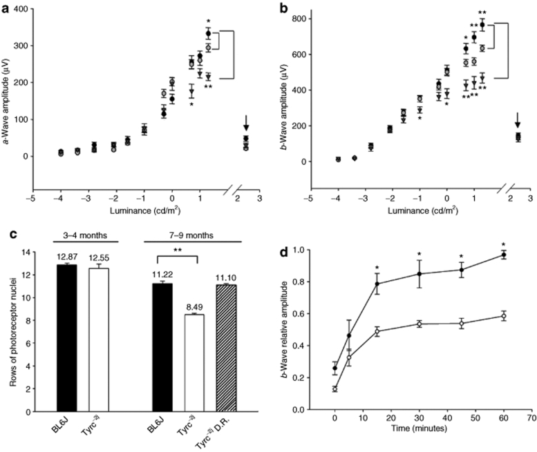
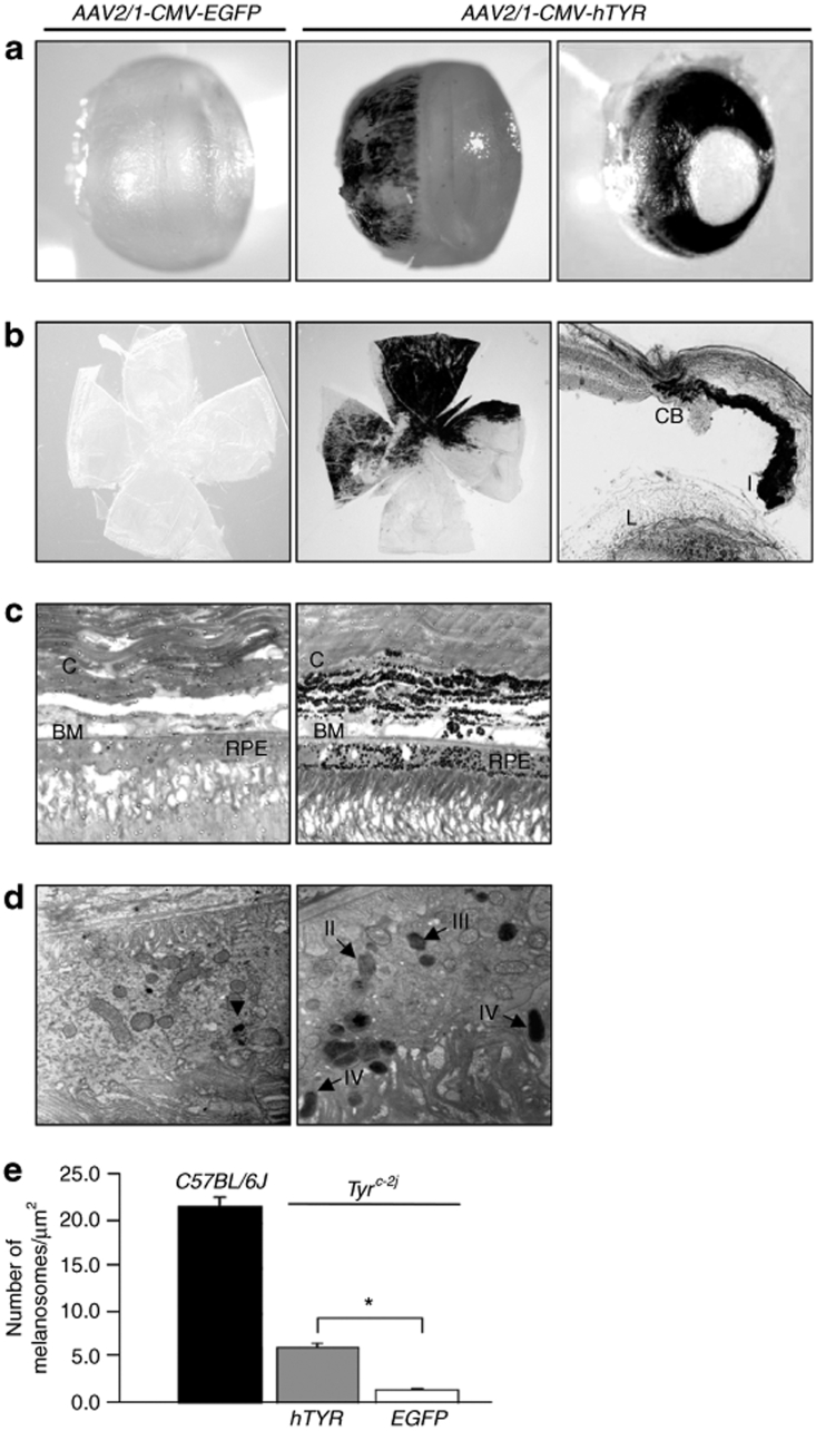
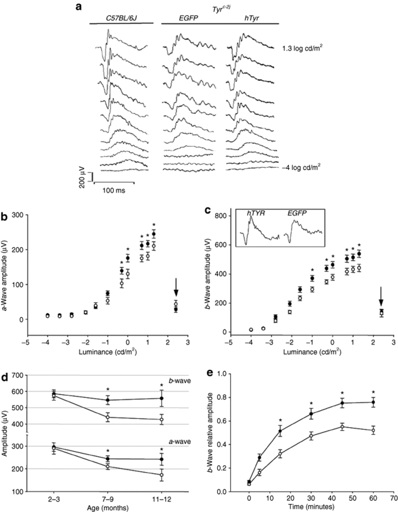
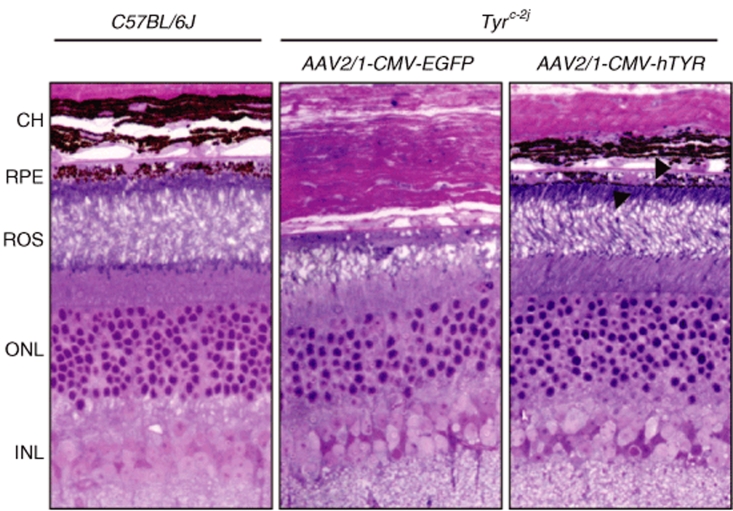
Oculo-cutaneous albinism type 1 (OCA1) is characterized by congenital hypopigmentation and is due to mutations in the TYROSINASE gene (TYR). In this study, we have characterized the morpho-functional consequences of the lack of tyrosinase activity in the spontaneous null mouse model of OCA1 (Tyr(c-2j)). Here, we show that adult Tyr(c-2j) mice have several retinal functional anomalies associated with photoreceptor loss. To test whether these anomalies are reversible upon TYR complementation, we performed intraocular administration of an adeno-associated virus (AAV)-based vector, encoding the human TYR gene, in adult Tyr(c-2j) mice. This resulted in melanosome biogenesis and ex novo synthesis of melanin in both neuroectodermally derived retinal pigment epithelium (RPE) and in neural crest-derived choroid and iris melanocytes. Ocular melanin accumulation prevented progressive photoreceptor degeneration and resulted in restoration of retinal function. Our results reveal novel properties of pigment cells and show that the developmental anomalies of albino mice are associated with defects occurring in postnatal life, adding novel insights on OCA1 disease pathogenesis. In addition, we provide proof-of-principle of an effective gene-based strategy relevant for future application in albino patients.


Figures

<b>Figure 1</b>
Figure 1
Characterization of Tyrc-2j mouse retinal function. (a) a- and (b) b-wave amplitudes under scotopic and photopic conditions in C57BL/6J pigmented and in C57BL/6J-c-2j (Tyrc-2j) albino mice. The amplitudes (mean ± SEM) evoked by increasing light intensities under scotopic conditions in 1- to 3-month-old (gray circles, n = 43 eyes) and 7- to 9-month-old (filled triangles, n = 14 eyes) Tyrc-2j mice, as well as 7- to 9-month-old controls (black circles, n = 30 eyes) are shown. Photopic conditions are indicated by an arrow. (c) Rows of photoreceptor nuclei in normal C57BL/6J and Tyrc-2j mice (black and white bars, respectively) at both 1–3 months (n = 4 eyes) and 7–9 months (pigmented n = 4; albino n = 7 eyes) of age. Tyrc-2j mice dark-reared (D.R.) since birth and analyzed at 9 months are represented in striped columns (n = 5 eyes). (d) Recovery of b-wave amplitude, relative to prebleach baseline levels (1 cd/m2/s), after bleaching (300 cd/m2 for 3 minutes) in 1-month-old Tyrc-2j (empty squares, n = 20 eyes) and wild-type age-match C57BL/6J mice (black circles, n = 6 eyes). Asterisks depict statistical significance (t-test, *P < 0.01; **P < 0.0003).
<b>Figure 2</b>
Figure 2
AAV2/1-mediated ocular gene transfer restores pigmentation in Tyrc-2j mice. (a) External appearance of Tyrc-2j mice eyes treated subretinally or in the posterior chamber at P30 and analyzed at 1 month of age. Left and middle panels show subretinal-injected eyes treated with AAV2/1-CMV-EGFP (left panel) and AAV2/1-CMV-hTYR (middle panel) vectors, respectively. The right panel shows Tyrc-2j mice eyes injected with AAV2/1-CMV-hTYR in the posterior chamber. (b) Flat-mounted retinas of 8-month-old Tyrc-2j mice treated subretinally at P30 with the control (left panel) and the therapeutic vectors (middle panel), respectively. The right panel shows the histological section of the iris of 8-month-old Tyrc-2j mice injected at 1 month with AAV2/1-CMV-hTYR injected in the posterior chamber. I, iris; CB, ciliary body; L, lens. Magnification ×20. (c) Semi-thin sections of 2-month-old Tyrc-2j treated and control mice retinas injected at P30 with AAV2/1-CMV-hTYR and AAV2/1-CMV-EGFP, respectively. C, choroids; BM, Bruch's membrane; RPE, retinal pigment epithelium. Magnification ×100. (d) Electron microscopy analysis of the RPE from 2-month-old Tyrc-2j mice injected subretinally at 1 month of age with AAV2/1-CMV-EGFP (left) and the contralateral eye with AAV2/1-CMV-hTYR (right). Black arrows indicate melanosomes at different stages of maturation (from stage I to IV) in the eye treated with AAV2/1-CMV-hTYR vector (right). The black arrowhead in the control retina (left) depicts a lipofuscin deposit. Magnification ×6,000. (e) Number of melanosomes at different stages of maturation in the RPE of C57BL/6J (black bar) and Tyrc-2j mice injected subretinally with AAV2/1-CMV-hTYR or AAV2/1-CMV-EGFP (gray and white bars, respectively; n = 3 eyes/group). Asterisks depict statistical significance (t-test, P < 0.05).
<b>Figure 3</b>
Figure 3
Retinal functional recovery following AAV2/1-CMV-hTYR treatment in Tyrc-2j mice. (a) Representative serial dark-adapted ERG recordings to increasing intensities of light stimuli in 2-month-old C57BL/6J pigmented animal (left) and Tyrc-2j mice treated with AAV2/1-CMV-EGFP (middle) or AAV2/1-CMV-hTYR (right) at one month of age. AAV2/1-CMV-hTYR treatment elicits b-waves responses at lower light intensities compared to AAV2/1-CMV-EGFP injected controls. The traces shown are the average of three responses for each light stimulus. (b) a- and (c) b-wave amplitudes (mean ± SEM) under scotopic and photopic (arrows) conditions recorded 6–8 months (7- to 9-month-old animals) after subretinal administration of AAV2/1 vectors harboring hTYR (black circles, n = 22) or EGFP (empty circles, n = 21) genes in Tyrc-2j mice. In the rectangle in C representative responses generated by 20 cd/m2/s light stimulus in a 7-month-old Tyrc-2j mouse treated with AAV2/1-CMV-hTYR (left) and AAV2/1-CMV-EGFP (right) at P30. (d) a- and b-wave maximum amplitudes in Tyrc-2j mice recorded 1–2, 6–8, and 10–11 months after treatment at P30 with AAV2/1-CMV-hTYR (black circles) and AAV2/1-CMV-EGFP (empty circles) vectors. (e) Recovery of b-wave amplitude in 2- to 3-month-old Tyrc-2j mice injected subretinally with AAV2/1-CMV-hTYR (black circles, n = 16) or AAV2/1-CMV-EGFP (empty triangles, n = 16) at P30. Asterisks depict statistical significance (t-test, P < 0.05).
<b>Figure 4</b>
Figure 4
Morphological analysis of Tyrc-2j retinas following AAV-mediated gene transfer. Semi-thin sections of 8 months old C57BL/6J (left) and Tyrc-2j mice treated at P30 with AAV2/1-CMV-EGFP (middle) and AAV2/1-CMV-hTYR (right). Arrowheads indicate the presence of melanin in the RPE and in choroid cells. CH, choroid; INL, inner nuclear layer; ONL, outer nuclear layer; ROS, rods outer segment; RPE, retinal pigment epithelium.

References

    1. Goding CR. Melanocytes: the new Black. Int J Biochem Cell Biol. 2007;39:275–279. - PubMed
    1. Marks MS., and , Seabra MC. The melanosome: membrane dynamics in black and white. Nat Rev Mol Cell Biol. 2001;2:738–748. - PubMed
    1. Lamb TD., and , Pugh EN., Jr Dark adaptation and the retinoid cycle of vision. Prog Retin Eye Res. 2004;23:307–380. - PubMed
    1. Thompson DA., and , Gal A. Vitamin A metabolism in the retinal pigment epithelium: genes, mutations, and diseases. Prog Retin Eye Res. 2003;22:683–703. - PubMed
    1. Lopes VS, Wasmeier C, Seabra MC., and , Futter CE. Melanosome maturation defect in Rab38-deficient retinal pigment epithelium results in instability of immature melanosomes during transient melanogenesis. Mol Biol Cell. 2007;18:3914–3927. - PMC - PubMed

Publication types

MeSH terms