

Angioid streaks, clinical course, complications, and current therapeutic management
- PMID: 19436620
- PMCID: PMC2697526
Angioid streaks, clinical course, complications, and current therapeutic management
Abstract
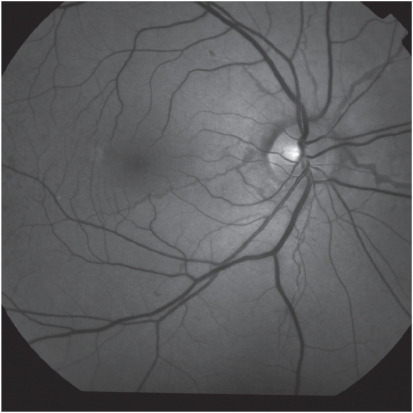
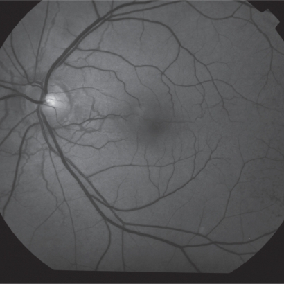
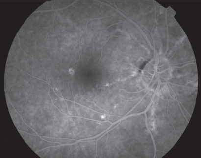
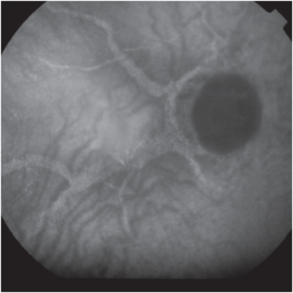
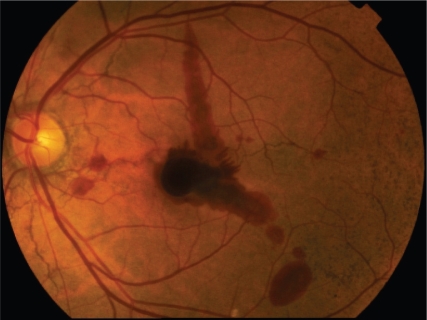
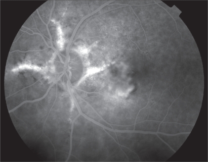
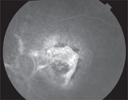
Angioid streaks are visible irregular crack-like dehiscences in Bruch's membrane that are associated with atrophic degeneration of the overlying retinal pigmented epithelium. Angioid streaks may be associated with pseudoxanthoma elasticum, Paget's disease, sickle-cell anemia, acromegaly, Ehlers-Danlos syndrome, and diabetes mellitus, but also appear in patients without any systemic disease. Patients with angioid streaks are generally asymptomatic, unless the lesions extend towards the foveola or develop complications such as traumatic Bruch's membrane rupture or macular choroidal neovascularization (CNV). The visual prognosis in patients with CNV secondary to angioid streaks if untreated, is poor and most treatment modalities, until recently, have failed to limit the devastating impact of CNV in central vision. However, it is likely that treatment with antivascular endothelial growth factor, especially in treatment-naive eyes to yield favorable results in the future and this has to be investigated in future studies.
Figures

References
-
- Doyne RW. Choroidal and retinal changes. The results of blows on the eyes. Trans Ophthalmol Soc U K. 1889;9:128.
-
- Knapp H. On the formation of dark angioid streaks as unusual metamorphosis of retinal hemorrhage. Arch Ophthalmol. 1892;26:289–292.
-
- Kofler A. Beitrage zur Kenntnis der angioid Streaks (Knapp) KIin Augenheilkd. 1917;82:134–149.
-
- Bock Z. KIinik und Anatomie der gefassahnlichen Streifen im Augenhintergrund. Z Augenheilkd. 1938;95:1–50.
-
- Hagedoorn A. Angioid streaks. Arch Ophthalmol. 1939;21:746–774. 935–965.
LinkOut - more resources
Full Text Sources
Other Literature Sources
