. 2009 May;5(5):e1000436.
doi: 10.1371/journal.ppat.1000436. Epub 2009 May 15.

The F(0)F(1)-ATP synthase complex contains novel subunits and is essential for procyclic Trypanosoma brucei

Affiliations

The F(0)F(1)-ATP synthase complex contains novel subunits and is essential for procyclic Trypanosoma brucei

Alena Zíková et al. PLoS Pathog. 2009 May.

Abstract

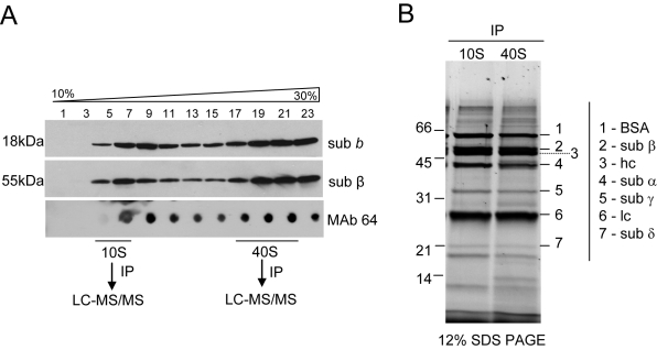
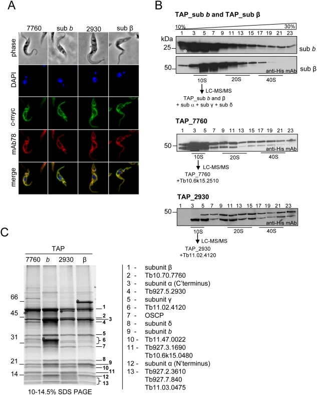
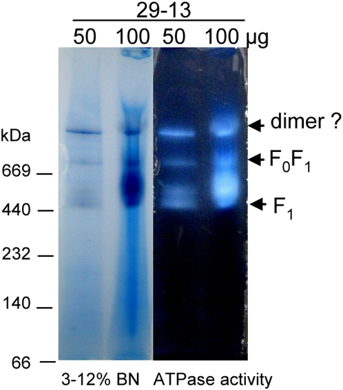
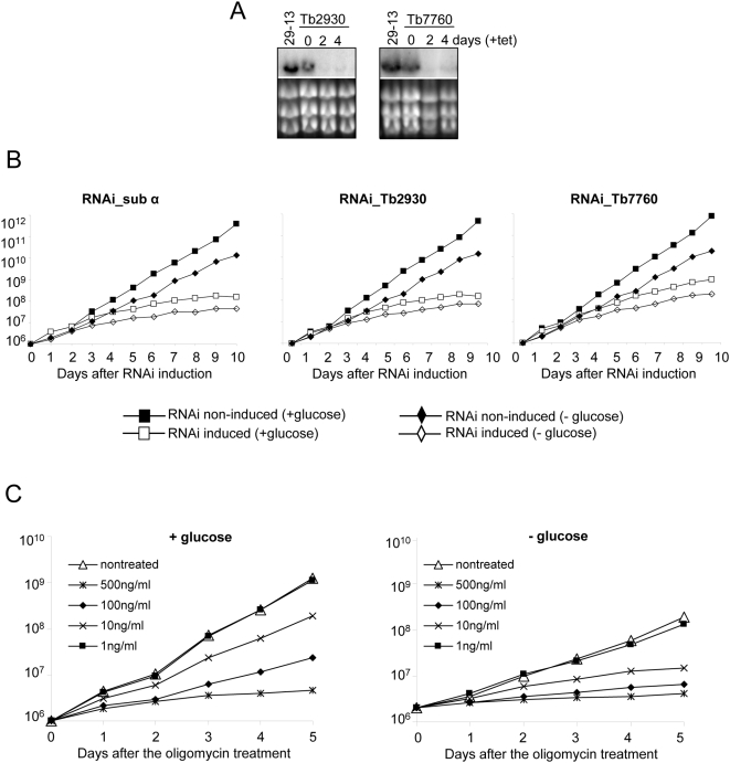
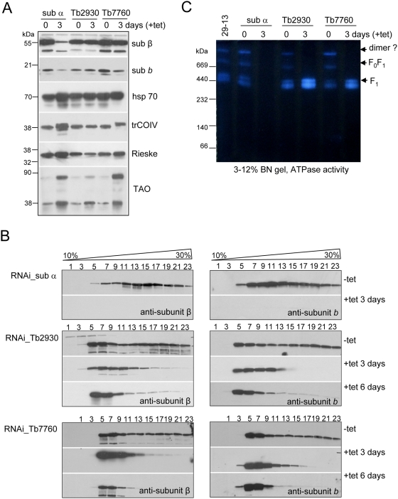
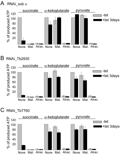
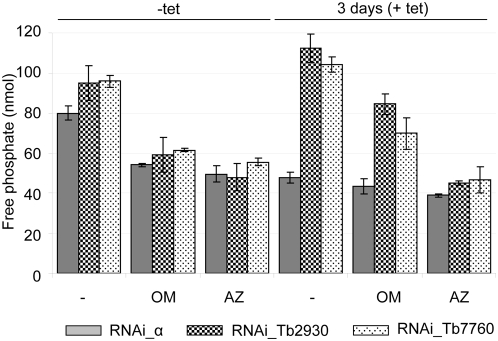
The mitochondrial F(0)F(1) ATP synthase is an essential multi-subunit protein complex in the vast majority of eukaryotes but little is known about its composition and role in Trypanosoma brucei, an early diverged eukaryotic pathogen. We purified the F(0)F(1) ATP synthase by a combination of affinity purification, immunoprecipitation and blue-native gel electrophoresis and characterized its composition and function. We identified 22 proteins of which five are related to F(1) subunits, three to F(0) subunits, and 14 which have no obvious homology to proteins outside the kinetoplastids. RNAi silencing of expression of the F(1) alpha subunit or either of the two novel proteins showed that they are each essential for the viability of procyclic (insect stage) cells and are important for the structural integrity of the F(0)F(1)-ATP synthase complex. We also observed a dramatic decrease in ATP production by oxidative phosphorylation after silencing expression of each of these proteins while substrate phosphorylation was not severely affected. Our procyclic T. brucei cells were sensitive to the ATP synthase inhibitor oligomycin even in the presence of glucose contrary to earlier reports. Hence, the two novel proteins appear essential for the structural organization of the functional complex and regulation of mitochondrial energy generation in these organisms is more complicated than previously thought.


Conflict of interest statement

The authors have declared that no competing interests exist.

Figures

Figure 1
Figure 1. Glycerol gradient fractionation and immunoprecipitation of F0F1-ATP synthase complex from T. brucei mitochondria.
(A) Western blot and native dot blot analyses of the 10–30% glycerol gradient-fractionated cleared mitochondrial lysate were performed using polyclonal antibodies against subunit b and β, and monoclonal antibody mAb64 to determine the sedimentation pattern of the F0F1-ATP synthase complex. MAb64 was further used to immunoprecipitate (IP) complexes from the 10S and 40S peaks and their protein compositions were analyzed by liquid-chromatography tandem mass spectrometry (LC-MS/MS). (B) Immunoprecipitated 10S and 40S complexes were fractionated on a 12% SDS PAGE gel and stained by Sypro Ruby. Protein bands corresponding to immunoglobulin heavy (hc) and light (lc) chains as well as predicted positions of F1 subunits α, β, γ and δ and the size standards are indicated.
Figure 2
Figure 2. Subcellular localization of tagged subunits and tandem-affinity purification (TAP) of F0F1-ATP synthase complex from T. brucei.
(A) Tagged subunits b, β, Tb7760 and Tb2930 of the F0F1-ATP synthase complex were visualized by fluorescence microscopy using polyclonal anti-c myc antiserum coupled with FITC-conjugated secondary antibody. Co-localization immunofluorescence was performed with monoclonal antibody mAb78 against the mt heat shock protein 70 . Top row, phase-contrast light microscopy of T. brucei cells; second row, 4,6-diamidino-2-phenylindole (DAPI)-staining of nuclear and kinetoplast DNA; third row, localization of tagged proteins; fourth row, staining of mitochondrial hsp70; bottom row, merged fluorescence images. (B) Fractionation of TAP_sub b, TAP_sub β, TAP_Tb2930 and TAP_Tb7760 TEV eluates on 10–30% glycerol gradients. Fractions were collected from the top of the gradients. Aliquots of odd-numbered fractions were analyzed by SDS-PAGE and probed with anti-His6 mAb. Positive fractions designated by a black line were further subjected to the second affinity purification step. Proteins identified in 10S complexes by LC-MS/MS are shown. The asterisk in the TAP_sub b panel indicates the position of the 25-kDa His-tagged TEV protease. The sizes of the protein marker are indicated on the left. (C) TAP_Tb7760, TAP_sub b, TAP_Tb2930 and TAP_sub β complexes were purified by Method 2, separated on a 10–14.5% polyacrylamide Tris-glycine gel and stained with Sypro Ruby. Numbers on the right indicate the results of MS analysis of individual bands. The asterisks indicate the tagged proteins. The sizes of the protein marker are indicated.
Figure 3
Figure 3. Analysis of the supramolecular organization of F0F1-ATP synthase complex from PF T. brucei.
Mt membranes from parental 29-13 cell line were solubilized by dodecyl maltoside and the mt complexes were separated on 3–12% BN PAGE. ATP synthase F1 particles, F0F1 monomer and putative dimer were identified by lead phosphate precipitates formed during the in-gel ATP hydrolysis assay followed by LC-MS/MS analysis of the corresponding gel bands. The sizes of the native high molecular weight marker (Amersham) are indicated.
Figure 4
Figure 4. Subunits α, Tb7760 and Tb2930 are important for the in vitro growth of procyclic parasites.
(A) Northern analyses of the corresponding mRNAs for Tb7760 and Tb2930 RNAi cell lines with the days sampled indicated; and stained gels of rRNAs in the lower panel serving as loading controls. (B) Growth curves of uninduced and induced RNAi-sub α (left), RNAi_Tb2930 (middle panel) and RNAi_Tb7760 (right) cell lines in the presence or absence of glucose. Cells were maintained in the exponential growth phase (between 106 and 107 cells/ml) and cumulative cell number represents the normalization of cell density by multiplication with the dilution factor. (C) Growth of the 29-13 procyclic cell line in the presence or absence of glucose and in response to treatment with the indicated concentrations of oligomycin. Cells were cultured and their growth measured as described for (B).
Figure 5
Figure 5. Effect of RNAi silencing of subunits α, Tb7760 or Tb2930 on steady-state abundance and integrity of F0F1-ATP synthase complex.
(A) The steady state abundance of examined proteins was analyzed by Western analysis of lysed crude mitochondria (7.5 µg of proteins per well) prepared from RNAi-induced cells after 3 days and from uninduced control cells. The blots were probed with antibodies against subunits β, b, trCOIV, Rieske protein, mt alternative oxidase (TAO) and mt hsp 70 as a loading control. (B) Western analyses of glycerol gradient fractions (numbered from top to bottom) from crude mitochondrial lysate of RNAi cell lines grown in the absence (−tet) or presence (+tet) of tetracycline for 3 and 6 days are shown. (C) In-gel ATP hydrolysis activity of T. brucei F0F1-ATP synthase complexes after ablation of subunit α, Tb7760 or Tb2930. Mitochondrial preparations were solubilized using dodecyl maltoside and separated by 3–12% BN PAGE. In-gel ATP hydrolysis/lead phosphate precipitation assay revealed bands representing putative monomeric and dimeric ATP synthase complexes and free F1 particles. The sizes of the native high molecular weight marker (Amersham) are indicated.
Figure 6
Figure 6. Effect of RNAi-mediated ablation of subunits α, Tb7760 or Tb2930 on mitochondrial ATP production.
Crude mitochondrial preparations from uninduced and 3-day RNAi-silenced RNA_sub α (A), RNAi_Tb2930 (B) and RNAi_Tb7760 (C) cells were obtained by digitonin extraction and ATP production in the three mitochondrial pathways was measured individually. ATP-production was triggered by the addition of ADP plus one of the following substrates, succinate, α-ketoglutarate and pyruvate/succinate. Uninduced cells (−tet) are shown in grey, induced (+tet) are shown in black. The tested substrate is indicated at the top. Malonate (Mal.), a specific inhibitor of succinate dehydrogenase, was used to inhibit ATP production by oxidative phosphorylation and atractyloside (Atrac.) was used to inhibit import of ADP into mitochondria. Addition of these compounds to the sample is indicated at the bottom of each panel. ATP production in mitochondria isolated from uninduced cells and tested without additions of malonate or atractyloside (None) was set to 100%. The bars represent means expressed as percentages from three independent RNAi inductions. Standard deviations are indicated.
Figure 7
Figure 7. Effect of RNAi-mediated ablation of subunits α, Tb7760 or Tb2930 on mitochondrial ATPase activity.
Crude mitochondrial preparations from uninduced and 3 days RNAi-silenced cells were obtained by digitonin extraction and ATPase activity was assayed by measuring release of free phosphate. Shown are data for RNAi_sub α (grey columns), RNAi_Tb2930 (small checker board columns) and RNAi_7760 (dotted diamonds columns) cell lines uninduced and induced for three days. ATP synthase inhibitors oligomycin (OM, 2.5 µg/ml) and azide (AZ, 1 mM) were added as indicated. Average numbers for three assays are shown, using extract preparation from three independent RNAi experiments.

Similar articles

Cited by

References

    1. Simarro PP, Jannin J, Cattand P. Eliminating human African trypanosomiasis: where do we stand and what comes next? PLoS Med. 2008;5:e55. doi:10.1371/journal.pmed.0050055. - PMC - PubMed
    1. Bringaud F, Riviere L, Coustou V. Energy metabolism of trypanosomatids: adaptation to available carbon sources. Mol Biochem Parasitol. 2006;149:1–9. - PubMed
    1. Besteiro S, Barrett MP, Riviere L, Bringaud F. Energy generation in insect stages of Trypanosoma brucei: metabolism in flux. Trends Parasitol. 2005;21:185–191. - PubMed
    1. Schnaufer A, Clark-Walker GD, Steinberg AG, Stuart K. The F(1)-ATP synthase complex in bloodstream stage trypanosomes has an unusual and essential function. EMBO J. 2005;24:4029–4040. - PMC - PubMed
    1. Brown SV, Hosking P, Li J, Williams N. ATP synthase is responsible for maintaining mitochondrial membrane potential in bloodstream form Trypanosoma brucei. Eukaryot Cell. 2006;5:45–53. - PMC - PubMed

Publication types

MeSH terms

LinkOut - more resources