. 2009;4(5):e5517.
doi: 10.1371/journal.pone.0005517. Epub 2009 May 13.

Attaching and effacing Escherichia coli downregulate DNA mismatch repair protein in vitro and are associated with colorectal adenocarcinomas in humans

Affiliations

Attaching and effacing Escherichia coli downregulate DNA mismatch repair protein in vitro and are associated with colorectal adenocarcinomas in humans

Oliver D K Maddocks et al. PLoS One. 2009.

Abstract

Background: Mucosa-associated Escherichia coli are frequently found in the colonic mucosa of patients with colorectal adenocarcinoma, but rarely in healthy controls. Chronic mucosal E. coli infection has therefore been linked to colonic tumourigenesis. E. coli strains carrying eae (encoding the bacterial adhesion protein intimin) attach intimately to the intestinal mucosa and are classed as attaching and effacing E. coli (AEEC). Enteropathogenic Escherichia coli (EPEC) are the most common form of AEEC identified in man. EPEC utilise a type III secretion system to translocate effector proteins into host cells and infection induces wide-ranging effects on the host cell proteome. We hypothesised that EPEC infection could influence molecular pathways involved in colorectal tumourigenesis.

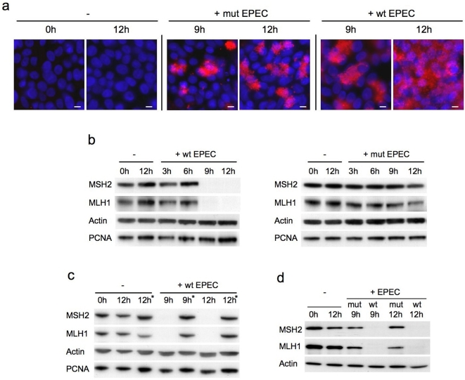
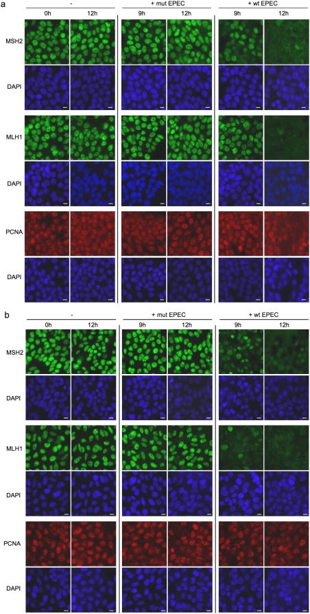
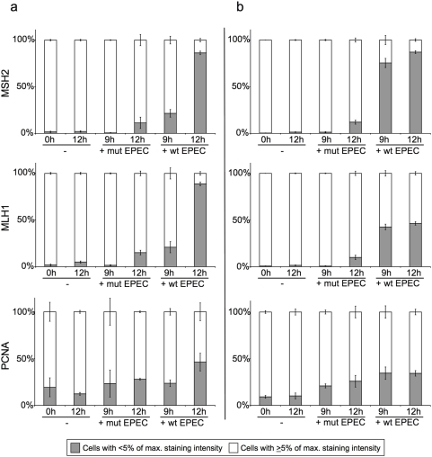
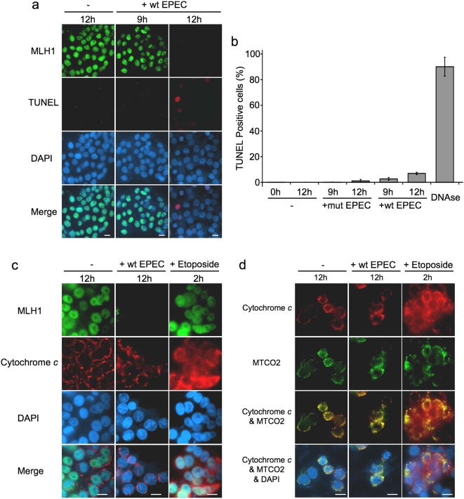
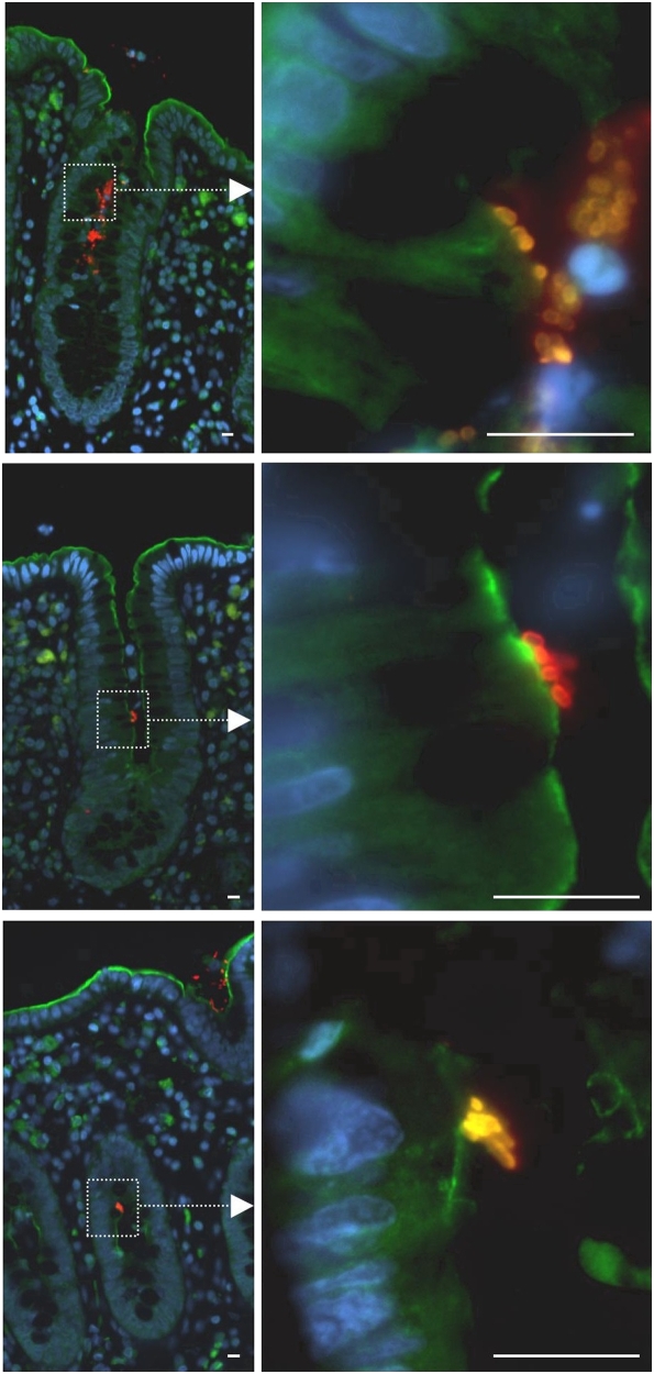
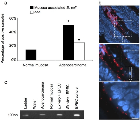
Methodology/principal findings: When co-cultured with human colorectal cell lines, EPEC dramatically downregulated the expression of key DNA mismatch repair proteins MSH2 and MLH1 in an attachment specific manner. Cytochrome c staining and TUNEL analysis confirmed that this effect was not a consequence of apoptosis/necrosis. Ex vivo human colonic mucosa was co-cultured with EPEC and probed by immunofluorescence to locate adherent bacteria. EPEC entered 10% of colonic crypts and adhered to crypt epithelial cells, often in the proliferative compartment. Adenocarcinoma and normal colonic mucosa from colorectal cancer patients (n = 20) was probed by immunofluorescence and PCR for AEEC. Mucosa-associated E. coli were found on 10/20 (50%) adenocarcinomas and 3/20 (15%) normal mucosa samples (P<0.05). AEEC were detected on 5/20 (25%) adenocarcinomas, but not normal mucosa samples (P<0.05).

Significance/conclusions: The ability of EPEC to downregulate DNA mismatch repair proteins represents a novel gene-environment interaction that could increase the susceptibility of colonic epithelial cells to mutations and therefore promote colonic tumourigenesis. The potential role of AEEC in colorectal tumourigenesis warrants further investigation.


Conflict of interest statement

Competing Interests: The authors have declared that no competing interests exist.

Figures

Figure 1
Figure 1. Co-culture of human colorectal cell lines with EPEC in vitro.
Immunofluorescence staining for E. coli (red) and DAPI nuclear staining (blue) shows the adherence patterns of wild-type (E2468/69) and mutant (UMD864) EPEC (a). Immunoblot analysis of DNA mismatch repair protein expression in human colorectal cancer cell lines co-cultured with EPEC: HT29 cells were either uninfected (−) or co-cultured with wild-type (+wt) or mutant (+mut) EPEC for 3–12 hours (b). HT29 cells were either uninfected or co-cultured with wild-type EPEC for 9 or 12 hours then treated with antibiotics for 36–39 hours (*) to remove infection (c). SW480 cells were either uninfected or co-cultured with wild-type or mutant EPEC for 9 or 12 hours (d). EPEC strain UMD864 does not express EspB, a protein required for a functional type III secretion system. Scale bar = 10 µm.
Figure 2
Figure 2. Immunofluorescence staining of DNA mismatch repair proteins in human colorectal cancer cell lines co-cultured with EPEC.
HT29 (a) and SW480 cells (b) were either uninfected (−) or co-cultured with wild-type (+wt) or mutant (+mut) EPEC for 9–12 hours. Fixed cells were probed for MSH2, MLH1 (both green) and PCNA (red) by immunofluorescence, nuclei were counterstained with DAPI (blue). Scale bar = 10 µm.
Figure 3
Figure 3. Quantitative image analysis of DNA mismatch repair protein immunostaining in human colorectal cancer cell lines co-cultured with EPEC.
HT29 (a) and SW480 cells (b) were either uninfected (−) or co-cultured with wild-type (+wt) or mutant (+mut) EPEC for 9–12 hours. Fixed cells were probed for MSH2, MLH1 and PCNA by immunofluorescence. Digital images were quantitatively analysed for nuclear staining intensity of the proteins of interest. According to staining intensity values, individual cells were categorised as having an intensity value greater than or equal to 5% of the maximum staining intensity (as measured in uninfected controls) or less than 5% of the maximum staining intensity. Cells with MSH2 or MLH1 staining below 5% of the maximum were classed as MMR protein deficient. The entire experiment was performed on three separate occasions for each cell line, error bars represent standard error of mean.
Figure 4
Figure 4. EPEC induced mismatch repair protein downregulation was not associated with apoptosis/necrosis.
HT29 cells either uninfected (−) or co-cultured with wild-type EPEC (+wt) for 9–12 hours were stained (red) using an in vitro cell death (TUNEL) detection kit, MLH1 was simultaneously stained (green) and nuclei were counterstained with DAPI (blue) (a). HT29 cells were either uninfected (−) or co-cultured with wild-type (+wt) or mutant (+mut) EPEC; TUNEL positive and negative cells were counted. Fixed HT29 cells treated with DNAase (to induce DNA strand breaks) were used as positive controls, error bars represent standard error of mean (n = 3) (b). HT29 cells were stained for MLH1 (green) and cytochrome c (red) (c) or for cytochrome c (red) and the mitochondrial marker MTCO2 (green) (d). Cells treated with etoposide for 2 h were positive controls for early apoptosis. Scale bar = 10 µm.
Figure 5
Figure 5. Co-culture of ex vivo adult human colonic mucosa with EPEC.
Sections of colonic mucosa were removed from surgical patients and co-cultured with wild-type EPEC for 12 hours, then fixed in buffered formalin and embedded in paraffin. Images show examples of tissue sections stained by immunofluorescence for E. coli (red) and ezrin (green) as a marker of the cell surface and DAPI (blue). Scale bar = 10 µm.
Figure 6
Figure 6. Analysis of human colorectal adenocarcinomas and normal colorectal tissue for attaching and effacing E. coli.
Paraffin embedded sections of adenocarcinoma and matched normal tissue from 20 patients were probed for E. coli by immunofluorescence and for the bacterial gene eae by PCR (*P<0.05, Fisher's Exact Test, two tails) (a). Example of an adenocarcinoma with mucosa associated E. coli (red), nuclei stained with DAPI (blue) (b) that tested positive for eae by PCR (c). Sections of ex vivo human colonic mucosa that were either co-cultured with (+) or without (−) wild-type EPEC were used as additional controls for PCR. Scale bar = 10 µm.

Similar articles

Cited by

References

    1. Stewart BW, Kleihues P, editors. World Cancer Report. Lyon: World health Organisation, International Agency for Research on Cancer, IARC Press; 2003. Colorectal Cancer. p. 198.
    1. de la Chapelle A. Genetic predisposition to colorectal cancer. Nat Rev Cancer. 2004;4:769–780. - PubMed
    1. Mager D. Bacteria and cancer: cause, coincidence or cure? A review. J Transl Med. 2006;4:14. - PMC - PubMed
    1. Martin HM, Campbell BJ, Hart CA, Mpofu C, Nayar M, et al. Enhanced Escherichia coli adherence and invasion in Crohn's disease and colon cancer. Gastroenterology. 2004;127:80–93. - PubMed
    1. Swidsinski A, Khilkin M, Kerjaschki D, Schreiber S, Ortner M, et al. Association between intraepithelial Escherichia coli and colorectal cancer. Gastroenterology. 1998;115:281–286. - PubMed

Publication types

MeSH terms

Substances