. 2009;4(5):e5544.
doi: 10.1371/journal.pone.0005544. Epub 2009 May 14.

Regulation of ErbB2 receptor status by the proteasomal DUB POH1

Affiliations

Regulation of ErbB2 receptor status by the proteasomal DUB POH1

Han Liu et al. PLoS One. 2009.

Abstract

Understanding the factors, which control ErbB2 and EGF receptor (EGFR) status in cells is likely to inform future therapeutic approaches directed at these potent oncogenes. ErbB2 is resistant to stimulus-induced degradation and high levels of over-expression can inhibit EGF receptor down-regulation. We now show that for HeLa cells expressing similar numbers of EGFR and ErbB2, EGFR down-regulation is efficient and insensitive to reduction of ErbB2 levels. Deubiquitinating enzymes (DUBs) may extend protein half-lives by rescuing ubiquitinated substrates from proteasomal degradation or from ubiquitin-dependent lysosomal sorting. Using a siRNA library directed at the full complement of human DUBs, we identified POH1 (also known as Rpn11 or PSMD14), a component of the proteasome lid, as a critical DUB controlling the apparent ErbB2 levels. Moreover, the effects on ErbB2 levels can be reproduced by administration of proteasomal inhibitors such as epoxomicin used at maximally tolerated doses. However, the extent of this apparent loss and specificity for ErbB2 versus EGFR could not be accounted for by changes in transcription or degradation rate. Further investigation revealed that cell surface ErbB2 levels are only mildly affected by POH1 knock-down and that the apparent loss can at least partially be explained by the accumulation of higher molecular weight ubiquitinated forms of ErbB2 that are detectable with an extracellular but not intracellular domain directed antibody. We propose that POH1 may deubiquitinate ErbB2 and that this activity is not necessarily coupled to proteasomal degradation.


Conflict of interest statement

Competing Interests: The authors have declared that no competing interests exist.

Figures

Figure 1
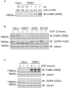
Figure 1. ErbB2 escapes EGF induced down-regulation.
A, Comparison of ErbB2 receptor levels in HeLa and SKBr3 cells. Cell lysate samples corresponding to the indicated number of cells were separated by SDS-PAGE and immunoblotted with ErbB2 antibodies and IR800-coupled secondary antibodies. The relative amount of ErbB2 per cell was calculated based on Odyssey scans as discussed in Materials and Methods. B, HeLa, HEK293T (H293T), A549, and DU145 cells were stimulated with 100 ng/ml EGF for 2 hours and lysed in parallel with unstimulated cells. The lysate was subjected to SDS-PAGE and immunoblotting with EGFR, ErbB2, and tubulin antibodies. EGFR is down-regulated after 2 hours stimulation, but ErbB2 remains stable. C, HeLa and SKBr3 cells were treated with 100 ng/ml EGF for 1 or 2 hours and analysed by immunoblotting with EGFR and ErbB2 antibodies. No EGFR was detected in SKBr3 cells. Note that EGFR antibody (2232) cross-reacts with high levels of ErbB2 in SKBr3 cells.
Figure 2
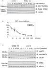
Figure 2. Effects of ErbB2 depletion on EGFR down-regulation and downstream signaling in HeLa cells.
A. HeLa cells were treated with ErbB2 siRNA or oligofectamine transfection reagent (control) for 48 hours before stimulation with 100 ng/ml EGF for various time periods. Lysates were analysed by immunoblotting with EGFR and ErbB2 antibodies. B. quantitation of A showing EGFR down-regulation was not significantly affected (data averaged from 3 experiments). C. HeLa cells were treated as in A and incubated with 10 ng/ml EGF for different time periods and lysed. Lysate was analysed by immunoblotting with ErbB2, pMAPK, MAPK, and tubulin antibodies. Levels of total MAPK and pMAPK were not affected by ErbB2 knock-down.
Figure 3
Figure 3. DUB screen for altered ErbB2 levels in HeLa cells.
siRNA mediated knockdown of DUBs was carried out in HeLa cells (siRNA at 40 nM). Cells were lysed after 72 hours and analysed by immunoblotting with ErbB2 antibody. A. The amount of ErbB2 for each sample was normalized to tubulin and expressed relative to control (non-targeting oligo) for each DUB contained in the library. B. ErbB2 levels in cells treated with a sub-set of the siGenome DUB library.
Figure 4
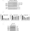
Figure 4. Multiple POH1 oligos down-regulate ErbB2.
A. siRNA mediated knockdown (KD) of candidates was repeated with four individual On Target Plus oligos incubated with HeLa cells for 48 hours. The results for POH1 were consistent. The knockdown of other candidates (MYSM1, USP14, BAP1, etc) with four individual oligos failed to show significant effects on ErbB2 levels (results from MYSM1 shown as an example). Lysates were also probed with EGFR, Met, and tubulin antibodies. The effect of knock-down of POH1 on EGFR and Met levels was much less pronounced than for ErbB2. B. quantitation of ErbB2, EGFR, and Met receptors for POH1 knockdown cells (averaged from 3 experiments). C. HeLa cells were treated with POH1 siRNA (four individual oligos) or oligofectamine transfection reagent (control) for 48 hours before lysis and analysed by immunoblotting for Transferrin receptor (TfR), Hrs, STAM, AMSH, and tubulin. Knock-down of POH1 showed minor effects on the levels of these proteins.
Figure 5
Figure 5. POH1 depletion does not differentially affect ErbB2 and EGFR transcription levels.
HeLa cells were treated±POH1 siRNA (two individual oligos) for 24 or 48 hours (left and right panels respectively) before RNA was extracted. Levels of mRNA of EGFR and ErbB2 were calculated relative to actin mRNA. Graph shows qRT-PCR results averaged from 3 experiments. Error bars show standard deviation.
Figure 6
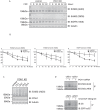
Figure 6. POH1 depletion and ErbB2 receptor turnover.
A, HeLa cells were treated±POH1 siRNA for 48 hours before incubation with 10 µg/ml cycloheximide. Cells were lysed and analysed by immunoblotting with ErbB2 29D8 and Ab20 antibodies, which recognize intracellular and extracellular epitopes of ErbB2 respectively, EGFR, and tubulin antibodies. B, quantitation shows that both EGFR (by antibody 1005) and ErbB2 (by antibodies Ab20 and 29D8) are turned over more rapidly in POH1 knock-down cells (data averaged from 3 experiments). C. HeLa cells were treated with four On Target Plus oligos (POH1) or with oligofectamine alone for 72 hours before lysis with hot lysis buffer. A higher molecular weight ErbB2 “smear” was observed in all 4 knock-down samples. D The high molecular weight smear associated with ErbB2 immuno-reactivity is sensitive to treatment with a deubiquitinase (USP2). HeLa cells were treated with POH1 siRNA or oligofectamine for 48 hours before lysis in the presence of NEM. ErbB2 was immunoprecipitated and treated in vitro with USP2 catalytic domain (100 nM, 8 hours, 37°C). Samples were analyzed by immunoblotting with ErbB2 antibodies targeting extracellular (Ab20) and intracellular (29D8) domains. Note that the smear detected with Ab20 is lost upon USP2 treatment whilst detection with the intracellular domain antibody increases. As a control for USP2 DUB-activity, EGFR was immunoprecipitated from EGF-stimulated (5 min) HeLa cells and treated in vitro with USP2 catalytic domain before SDS-PAGE and western blotting with anti-Ubiquitin.
Figure 7
Figure 7. Cell surface levels of ErbB2 are relatively insensitive to POH1 knock-down.
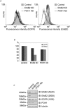
A, HeLa cells were treated with ErbB2, POH1 siRNA or oligofectamine alone for 48 hours before detachment with 2 mM EDTA. One million cells from each condition were labelled with FITC-conjugated ErbB2 and phycoerythrin (PE) conjugated EGFR antibodies and then analysed by flow cytometry. B, quantification of A shows relative amounts of fluorescence for each condition. C, samples from A were analysed by immunoblotting with ErbB2 (Ab20 and 29D8), EGFR, POH1, and tubulin antibodies.
Figure 8
Figure 8. POH1 deletion increases levels of free ubiquitin and ubiquitinated proteins.
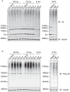
HeLa cells were treated with POH1 siRNA (oligo 1–4) or control reagent for 48 or 72 hours before lysis with “hot lysis” SDS-buffer. Equal amounts of cell lysates were analysed by immunoblotting with A, polyclonal anti-ubiquitin (recognising ubiquitinated proteins and free ubiquitin) and B, FK1 monoclonal anti-ubiquitin (recognising only polyubiquitinated proteins). As with proteasome inhibitor treatment (Lact: lactacystin, 10 µM and Epo: epoxomicin, 10 µM or 1 µM left and right panels respectively), POH1 depletion caused an accumulation of ubiquitinated proteins in the cell, but in contrast to the inhibitors, which deplete free ubiquitin, levels of free ubiquitin were elevated.
Figure 9
Figure 9. Chronic treatment with a proteasome inhibitor replicates effects of POH1 depletion on ErbB2.
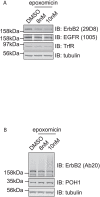
HeLa cells were incubated with epoxomicin (8 nM or 10 nM) or DMSO for 48 hours (fresh inhibitors were applied at 24 hours). Cell lysates obtained by “hot lysis” were subjected to SDS-PAGE followed by immunoblotting with ErbB2, EGFR, TrfR, POH1, and tubulin antibodies. Epoxomicin treatment resulted in the apparent loss of ErbB2 and concomitant appearance of a high molecular weight smear.

Similar articles

Cited by

References

    1. Yarden Y, Sliwkowski MX. Untangling the ErbB signalling network. Nat Rev Mol Cell Biol. 2001;2:127–137. - PubMed
    1. Sorkin A, Goh LK. Endocytosis and intracellular trafficking of ErbBs. Exp Cell Res. 2008;314:3093–3106. - PMC - PubMed
    1. Urbe S. Ubiquitin and endocytic protein sorting. Essays Biochem. 2005;41:81–98. - PubMed
    1. Lenferink AE, Pinkas-Kramarski R, van de Poll ML, van Vugt MJ, Klapper LN, et al. Differential endocytic routing of homo- and hetero-dimeric ErbB tyrosine kinases confers signaling superiority to receptor heterodimers. Embo J. 1998;17:3385–3397. - PMC - PubMed
    1. Wang Z, Zhang L, Yeung TK, Chen X. Endocytosis deficiency of epidermal growth factor (EGF) receptor-ErbB2 heterodimers in response to EGF stimulation. Mol Biol Cell. 1999;10:1621–1636. - PMC - PubMed

Publication types