. 2009 Nov-Dec;22(6):425-36.
doi: 10.1002/jmr.957.

Developing bifunctional beta-lactamase molecules with built-in target-recognizing module for prodrug therapy: identification of Enterobacter Cloacae P99 cephalosporinase loops suitable for randomization and phage-display selection

Affiliations

Developing bifunctional beta-lactamase molecules with built-in target-recognizing module for prodrug therapy: identification of Enterobacter Cloacae P99 cephalosporinase loops suitable for randomization and phage-display selection

Girja S Shukla et al. J Mol Recognit. 2009 Nov-Dec.

Abstract

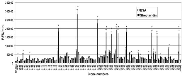
This study was focused on developing catalytically active beta-lactamase enzyme molecules that have target-recognizing sites built within their scaffold. Using phage-display approach, nine libraries were constructed by inserting the randomized linear or cysteine-constrained heptapeptides in the five different loops on the outer surface of P99 beta-lactamase molecule. The pIII signal peptide of Sec-pathway was employed for a periplasmic translocation of the beta-lactamase fusion protein, which we found more efficient than the DsbA signal peptide of SRP-pathway. The randomized heptapeptide loops replaced native amino acids between positions (34)Y-(37)K, (238)M-(246)A, (275)N-(280)A, (305)A-(311)S, or (329)I-(334)I of the P99 beta-lactamase molecules for generating the loop-1 to -5 libraries, respectively. The diversity of each loop library was judged by counting the primary and beta-lactamase-active clones. The linear peptide inserts in the loop-2 library showed the maximum number of the beta-lactamase-active clones, followed by the loop-5, loop-3, and loop-4. The insertion of the cysteine-constrained loops exhibited a dramatic loss of the enzyme-active beta-lactamase clones. The complexity of the loop-2 linear library, as determined by the frequency and diversity of amino acid distributions in the randomized region, appears consistent with the standards of other types of phage display library systems. The selection of the loop-2 linear library on streptavidin protein as a test target identified several beta-lactamase clones that specifically bound to streptavidin. In conclusion, this study identified the suitability of the loop-2 of P99 beta-lactamase for constructing a phage-display library of the beta-lactamase enzyme-active molecules that can be selected against a target. This is an enabling step in our long-term goal of developing bifunctional beta-lactamase molecules against cancer-specific targets for enzyme prodrug therapy of cancer.


Figures

Figure 1
Figure 1
Randomized loops of β-lactamase. (A) Schematic representation of different loops (solid black) of the Enterobacter cloacae P99 cephalosporinase molecule, based on the published X-ray crystallographic structure (Lobkovsky et al., 1994), that were selected for randomization. (B) The Molscript drawing of surface structure of the E cloacae P99 cephalosporinase molecule showing that all the selected loops (black) are on one face of the outer surface (based on PDB, 1BLS).
Figure 2
Figure 2
Relevant portions of synthetic E. cloacae P99 cephalosporinase molecule showing native amino acid sequences that were replaced by random linear or cysteine-constrained heptapeptide inserts in different loops and also the mutations that resulted from the creation of cloning sites. The amino acid sequence of wild-type β-lactamase is based on the Protein Data Bank entry (1BLS). The amino acids which were replaced for construction of different loop libraries are highlighted and written in white. The mutated amino acids in synthetic β-lactamase molecules are highlighted and written in black. Amino acids are presented as single-letter codes.
Figure 3
Figure 3
Schematic presentation of phagemid vector design and features. The vector has the modified E. cloacae P99 cephalosporinase (BLA) gene that allows cloning of random peptide sequences in the different loops of the BLA molecule. Five random sites presented in the BLA gene correspond to the insertion sites of (nnk)7 or tgt(nnk)7tgc for the linear or the cysteine-constrained heptapeptides, respectively. The vector sequences were substituted at 356–3361 bp for loop-1, 3968–3988 bp for loop-2, 4079–4090 bp for loop-3, 4169–4183 bp for loop-4, or 4241–4252 bp for loop-5 library. CAT = Chloramphenicol acetyltransferase.
Figure 4
Figure 4
Western blot analysis of phage proteins using anti-pIII antibody. (A) Immunodetection of SDS–PAGE-separated phage proteins (~1 × 10¹⁰ phage/well, proteins normalized). Lane 1 represents the phage from vector using the pIII signal sequence (pIIIss) for the pIII-β-lactamase fusion translocation. Lane 2 represents phage from vector using the DsbA signal sequence (DsbAss) for the pIII-β-lactamase fusion translocation. (B) Protein staining of the membrane that was used for immunodetection of pIII (4A above). An equal gel loading and membrane-transfer of the proteins are evidenced by equal intensities of pVIII and other visible bands of phage proteins in the two lanes. (C) The data from densitometric analyses of the bands are plotted as the percentage of the total pIII molecules that present the β-lactamase as fusion protein. Each bar represents mean ± SE of three analyses. *The significance of difference between the groups was evaluated by Student’s t-test; p ≤ 0.05 was considered to be significant. White bar = phage from vector with pIIIss; black bar = phage from vector with DsbAss.
Figure 5
Figure 5
Screening of β-lactamase clones selected from the loop-2 linear library panning for their binding to streptavidin (target) and BSA (control) proteins. The bars represent the mean ± SE of the two experiments done in triplicate. The binding was assessed by measuring the residual β-lactamase activity using Fluorocillin Green substrate. *The significance of differences between streptavidin and BSA bindings were evaluated by Student’s t-test; p ≤ 0.05 was considered to be significant. Clone numbers represent identity of the selected clones. NR-BLA = the lysate from bacteria transformed with the vector expressing the non-randomized β-lactamase loops; Library = the lysate from the unselected bacterial loop-2 linear library; RFU = relative fluorescence unit.

References

    1. Bagshawe KD. Antibody-directed enzyme prodrug therapy (ADEPT) for cancer. Expert Rev Anticancer Ther. 2006;6:1421–1431. - PubMed
    1. Bhatia J, Sharma SK, Chester KA, Pedley RB, Boden RW, Read DA, Boxer GM, Michael NP, Begent RH. Catalytic activity of an in vivo tumor targeted anti-CEA scFv::carboxypeptidase G2 fusion protein. Int J Cancer. 2000;85:571–577. - PubMed
    1. Cantu C, 3rd, Huang W, Palzkill T. Cephalosporin substrate specificity determinants of TEM-1 beta-lactamase. J Biol Chem. 1997;272:29144–29150. - PubMed
    1. Cartwright SJ, Waley SG. Purification of beta-lactamases by affinity chromatography on phenylboronic acid-agarose. Biochem J. 1984;221:505–512. - PMC - PubMed
    1. Cortez-Retamozo V, Backmann N, Senter PD, Wernery U, De Baetselier P, Muyldermans S, Revets H. Efficient cancer therapy with a nanobody-based conjugate. Cancer Res. 2004;64:2853–2857. - PubMed

Publication types

LinkOut - more resources