

Ectopic growth of hippocampal mossy fibers in a mutated GAP-43 transgenic mouse with impaired spatial memory retention
- PMID: 19437419
- PMCID: PMC2801775
- DOI: 10.1002/hipo.20635
Ectopic growth of hippocampal mossy fibers in a mutated GAP-43 transgenic mouse with impaired spatial memory retention
Abstract
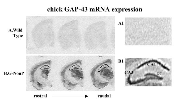
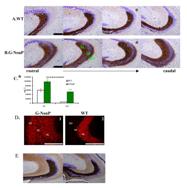
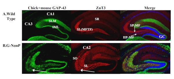
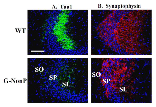
In a previous study, it was shown that transgenic mice, designated G-NonP, forget the location of a water maze hidden platform when tested 7 days after the last training day (Holahan and Routtenberg (2008) Hippocampus 18:1099-1102). The memory loss in G-NonP mice might be related to altered hippocampal architecture suggested by the fact that in the rat, 7 days after water maze training, there is discernible mossy fiber (MF) growth (Holahan et al. (2006) Hippocampus 16:560-570; Rekart et al. (2007) Learn Mem 14:416-421). In the present report, we studied the distribution of the MF system within the hippocampus of naïve, untrained, G-NonP mouse. In WT mice, the MF projection was restricted to the stratum lucidum of CA3 with no detectable MF innervation in distal stratum oriens (dSO). In G-NonP mice, in contrast, there was an ectopic projection terminating in the CA3 dSO. Unexpectedly, there was nearly a complete loss of immunostaining for the axonal marker Tau1 in the G-NonP transgenic mice in the MF terminal fields indicating that transgenesis itself leads to off-target consequences (Routtenberg (1996) Trends Neurosci 19:471-472). Because transgenic mice overexpressing nonmutated, wild type GAP-43 do not show this ectopic growth (Rekart et al., in press) and the G-NonP mice overexpress a mutated form of GAP-43 precluding its phosphorylation by protein kinase C (PKC), the possibility exists that permanently dephosphorylated GAP-43 disrupts normal axonal fasciculation which gives rise to the ectopic growth into dSO.
Figures
Comment in
-
Adult learning and remodeling of hippocampal mossy fibers: unheralded participant in circuitry for long-lasting spatial memory.Hippocampus. 2010 Jan;20(1):44-5. doi: 10.1002/hipo.20664. Hippocampus. 2010. PMID: 19554645
References
-
- Aarts LH, Verkade P, van Dalen JJ, van Rozen AJ, Gispen WH, Schrama LH, Schotman P. B-50/GAP-43 potentiates cytoskeletal reorganization in raft domains. Mol Cell Neurosci. 1999;14(2):85–97. - PubMed
-
- Aigner L, Arber S, Kapfhammer JP, Laux T, Schneider C, Botteri F, Brenner H-R, Caroni P. Overexpression of the neural growth-associated protein GAP-43 induces nerve sprouting in the adult nervous system of transgenic mice. Cell. 1995;83:269–278. - PubMed
-
- Alexander KA, Wakim BT, Doyle GS, Walsh KA, Storm DR. Identification and characterization of the calmodulin-binding domain of neuromodulin, a neurospecific calmodulin-binding protein. Journal of Biological Chemistry. 1988;263(16):7544–7549. - PubMed
-
- Baizer L, Alkan S, Stocker K, Ciment G. Chicken growth-associated protein (GAP)-43: primary structure and regulated expression of mRNA during embryogenesis. Brain Res Mol Brain Res. 1990;7(1):61–8. - PubMed
-
- Basi GS, Jacobson RD, Virag I, Schilling J, Skene JH. Primary structure and transcriptional regulation of GAP-43, a protein associated with nerve growth. Cell. 1987;49(6):785–791. - PubMed
Publication types
MeSH terms
Substances
Grants and funding
LinkOut - more resources
Full Text Sources
Medical
Molecular Biology Databases
Miscellaneous
