. 2009 May;214(5):645-54.
doi: 10.1111/j.1469-7580.2009.01071.x.

Coexistence of adrenergic and cholinergic nerves in the inferior hypogastric plexus: anatomical and immunohistochemical study with 3D reconstruction in human male fetus

Affiliations

Coexistence of adrenergic and cholinergic nerves in the inferior hypogastric plexus: anatomical and immunohistochemical study with 3D reconstruction in human male fetus

Bayan Alsaid et al. J Anat. 2009 May.

Abstract

Classic anatomical methods have failed to determine the precise location, origin and nature of nerve fibres in the inferior hypogastric plexus (IHP). The purpose of this study was to identify the location and nature (adrenergic and/or cholinergic) of IHP nerve fibres and to provide a three-dimensional (3D) representation of pelvic nerves and their relationship to other anatomical structures. Serial transverse sections of the pelvic portion of two human male fetuses (16 and 17 weeks' gestation) were studied histologically and immunohistochemically, digitized and reconstructed three-dimensionally. 3D reconstruction allowed a 'computer-assisted dissection', identifying the precise location and distribution of the pelvic nerve elements. Proximal (supra-levator) and distal (infra-levator) communications between the pudendal nerve and IHP were observed. By determining the nature of the nerve fibres using immunostaining, we were able to demonstrate that the hypogastric nerves and pelvic splanchnic nerves, which are classically considered purely sympathetic and parasympathetic, respectively, contain both adrenergic and cholinergic nerve fibres. The pelvic autonomic nervous system is more complex than previously thought, as adrenergic and cholinergic fibres were found to co-exist in both 'sympathetic' and 'parasympathetic' nerves. This study is the first step to a 3D cartography of neurotransmitter distribution which could help in the selection of molecules to be used in the treatment of incontinence, erectile dysfunction and ejaculatory disorders.


Figures

Fig. 1
Fig. 1
Transverse sections thought the pelvis of 17-week-old male fetus (A) and of 16-week-old male fetus (E), at ureterovesical junction level, stained with HES and scanned at 4800 dots/inch (dpi) optical resolution, (B) Microscopic aspect (×20) of pelvic splanchnic nerve (frame in A) immuno-labelled with anti-S100, (C,D) same nerve shows positivity to anti-TH and to anti-VAChT. (F) Microscopic aspect (×40) of right hypogastric nerve (frame in E) immuno-labelled with anti-S100, (G) nerve positive to anti-TH and (H) some nerve fibres are also positive to anti VAChT (IIA: Internal Iliac Artery, LST: Lumbo-Sacral Trunk, PSN: Pelvic Splanchnic nerve, RHN: Right Hypogastric nerve, S2: Second Sacral root, S3: Third Sacral root).
Fig. 2
Fig. 2
Two serial transverse sections of 17-week-old male fetus (200 µm interval), immuno-stained with anti-S100 and scanned at optical resolution of 4800 dpi. Nervous fibres leave the pudendal nerve (A, head arrow) to reach the inferior hypogastric plexus via the lateral face of its posterior portion (B, head arrow) (IHP: Inferior Hypogastric Plexus, OIM: Obturator Internal Muscle, PM: Piriform Muscle, PN: Pudendal Nerve, SN: Sciatic nerve).
Fig. 3
Fig. 3
Serial transverse sections (5 µm thickness) of 17-week-old male fetus (bladder neck level). (A) Reference section stained with HES, scanned at optical resolution of 4800 dpi, (B) Microscopic aspect (×10) of the inferior hypogastric plexus (frame in A), nervous fibres and ganglia immuno-stained with anti-S100, fibres and ganglia positive to the anti-VAChT (C – black arrow) and/or positive to anti-TH (D – white arrow) (IHP: Inferior Hypogastric Plexus, VD: Vas Deferens).
Fig. 4
Fig. 4
(A) Transverse scanned section of 16-week-old male fetus (urethral sphincter level), immuno-stained with anti-S100, (B) microscopic aspect (×10) of lateral side of urethral sphincter (left frame in A), fibres from the cavernous nerves (CN) destined to the corpus cavernosum were positive for anti-VAChT, (C) some fibres shows positivity with anti-TH, (D,E) microscopic aspects (×10) of dorsal nerve of the penis (DPN) (right frame in A) show positivity to the two immuno-markers.
Fig. 5
Fig. 5
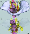
Computer assisted 3-dimensional reconstruction of a 17-week-old male fetal pelvis, (A) global view of the pelvis, (B) view with transparency of bones and muscles (A: Aorta, B: Bladder, CC: Corpus Cavernosum, CIA: Common Iliac Artery, DNP: Dorsal Nerve of the Penis, EIA: External Iliac Artery, FN: Fibular Nerve, IHP: Inferior Hypogastric Plexus, L5: 5th Lumbar root, LHN: Left Hypogastric Nerve, ON: Obturator Nerve, PD: Pelvic Diaphragm, PN: Pudendal Nerve, P: Prostate, R: Rectum, S1: 1st Sacral root, S2: 2nd Sacral root, S3: 3rd Sacral root, SHP: Superior Hypogastric Plexus, ST: Sympathetic Trunk, TN: Tibial Nerve, U: Ureter, UM: Umbilical Artery).
Fig. 7
Fig. 7
Computer assisted 3-D reconstruction of a 17-week-old male fetal pelvis. (A) Close lateral view with transparency of the inferior hypogastric plexus nervous fibres, quadrangular shape of plexus in white quadrangle, adrenergic fibres (in green) and cholinergic fibres (in purple), hypogastric nerves and pelvic splanchnic nerves contain both fibres’ types, adrenergic fibres mostly situated in the superior portion of the plexus, cholinergic fibres intend to concentrate in the inferior portion. (B, C) Posterior and lateral views of the prostate and seminal vesicles with nervous fibres transparency. The fibres at the proximal seminal vesicles portion follow two courses: posterior and lateral. The majority of adrenergic fibres distributing in the bladder neck, the seminal vesicles, the vas deferens and the prostate; the cholinergic fibres innerve the same structures and continue to reach the erectile bodies (star in C) (AF: Adrenergic Fibres, B: Bladder, CC: Corpus Cavernosum, CF: Cholinergic Fibres, CN: Cavernous Nerve, CS: Corpus Spongiosum, IHP: Inferior Hypogastric Plexus, LHN: Left Hypogastric Nerve, P: Prostate, PSN: Pelvic Splanchnic Nerve, R: Rectum, SV: Seminal Vesicle, U: Ureter, UVB: Ureterovesical Branches, VD: Vas Deferens).
Fig. 6
Fig. 6
Computer assisted 3-D reconstruction of a 17-week-old male fetal pelvis, antero-lateral view of the pelvic organs without bones and with muscles transparency. Hypogastric nerves and pelvic splanchnic nerves join the inferior hypogastric plexus at the level of the superior angle and the posterior edge, proximal (supra-levator) and distal (infra-levator) communications between the pudendal nerve and the plexus were observed (B: Bladder, CC: Corpus Cavernosum, CN: Cavernous Nerve, CS: Corpus Spongiosum, DC: Distal Communication, DNP: Dorsal Nerve of the Penis, IHP: Inferior Hypogastric Plexus, HN: Hypogastric Nerve, P: Prostate, PC: Proximal Communication, PD: Pelvic Diaphragm, PN: Pudendal Nerve, PSN: Pelvic Splanchnic Nerve, SHP: Superior Hypogastric Plexus, R: Rectum, U: Ureter, UVB: Ureterovesical Branches).

References

    1. Arango-Toro O, Domenech-Mateu JM. Development of the pelvic plexus in human embryos and fetuses and its relationship with the pelvic viscera. Eur J Morphol. 1993;31:193–208. - PubMed
    1. Arango Toro O, Domenech Mateu JM. [Anatomic and clinical evidence of intrapelvic pudendal nerve and its relation with striated sphincter of the urethra] Acta Urol Esp. 2000;24:248–254. - PubMed
    1. Baader B, Herrmann M. Topography of the pelvic autonomic nervous system and its potential impact on surgical intervention in the pelvis. Clin Anat. 2003;16:119–130. - PubMed
    1. Baljet B, Drukker J. Some aspects of the innervation of the abdominal and pelvic organs in the human female fetus. Acta Anat (Basel) 1982;111:222–230. - PubMed
    1. Benoît G, Droupy S, Quillard J, Paradis V, Giuliano F. Supra and infralevator neurovascular pathways to the penile corpora cavernosa. J Anat. 1999;195:605–615. Pt 4. - PMC - PubMed

MeSH terms

LinkOut - more resources