

The dynamic cilium in human diseases
- PMID: 19439065
- PMCID: PMC2694804
- DOI: 10.1186/1755-8417-2-3
The dynamic cilium in human diseases
Abstract
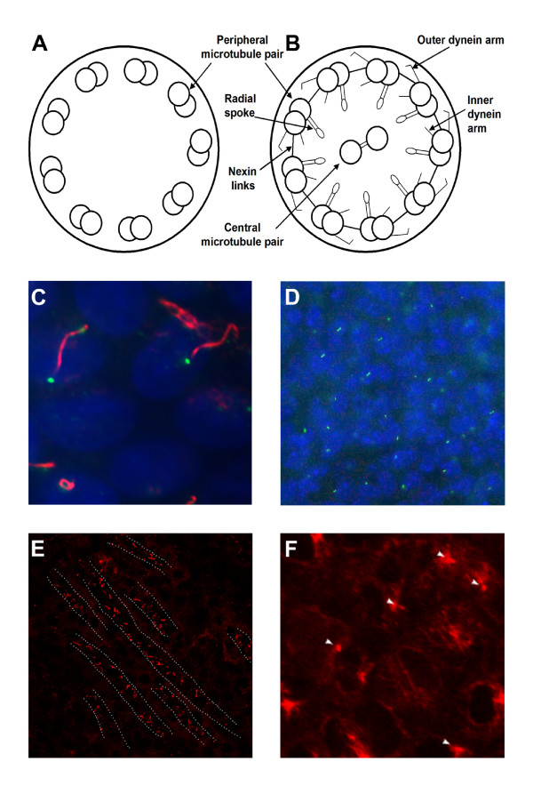
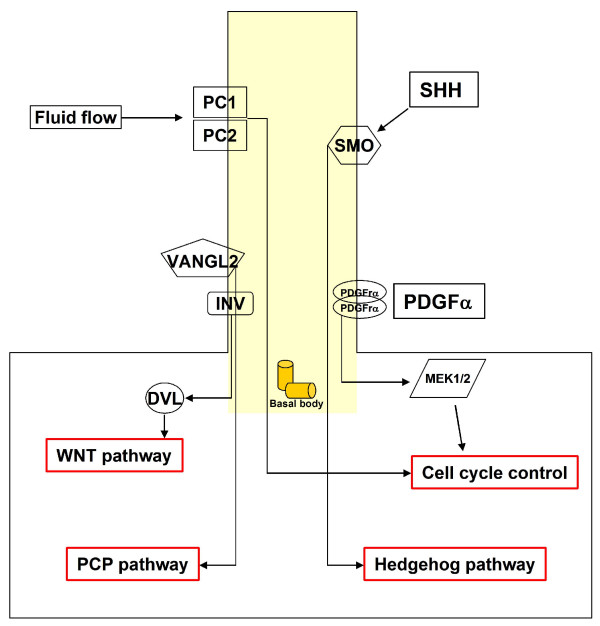
Cilia are specialized organelles protruding from the cell surface of almost all mammalian cells. They consist of a basal body, composed of two centrioles, and a protruding body, named the axoneme. Although the basic structure of all cilia is the same, numerous differences emerge in different cell types, suggesting diverse functions. In recent years many studies have elucidated the function of 9+0 primary cilia. The primary cilium acts as an antenna for the cell, and several important pathways such as Hedgehog, Wnt and planar cell polarity (PCP) are transduced through it. Many studies on animal models have revealed that during embryogenesis the primary cilium has an essential role in defining the correct patterning of the body. Cilia are composed of hundreds of proteins and the impairment or dysfunction of one protein alone can cause complete loss of cilia or the formation of abnormal cilia. Mutations in ciliary proteins cause ciliopathies which can affect many organs at different levels of severity and are characterized by a wide spectrum of phenotypes. Ciliary proteins can be mutated in more than one ciliopathy, suggesting an interaction between proteins. To date, little is known about the role of primary cilia in adult life and it is tempting to speculate about their role in the maintenance of adult organs. The state of the art in primary cilia studies reveals a very intricate role. Analysis of cilia-related pathways and of the different clinical phenotypes of ciliopathies helps to shed light on the function of these sophisticated organelles. The aim of this review is to evaluate the recent advances in cilia function and the molecular mechanisms at the basis of their activity.
Figures
References
-
- Satir P, Christensen ST. Overview of structure and function of mammalian cilia. Annu Rev Physiol. 2007;69:377–400. - PubMed
-
- Basu B, Brueckner M. Cilia multifunctional organelles at the center of vertebrate left-right asymmetry. Curr Top Dev Biol. 2008;85:151–174. - PubMed
-
- Nonaka S, Tanaka Y, Okada Y, Takeda S, Harada A, Kanai Y, Kido M, Hirokawa N. Randomization of left-right asymmetry due to loss of nodal cilia generating leftward flow of extraembryonic fluid in mice lacking KIF3B motor protein. Cell. 1998;95:829–837. - PubMed
-
- Menco BP. Ultrastructural aspects of olfactory transduction and perireceptor events. Semin Cell Biol. 1994;5:11–24. - PubMed
-
- Yamamoto M, Kataoka K. Electron microscopic observation of the primary cilium in the pancreatic islets. Arch Histol Jpn. 1986;49:449–457. - PubMed
LinkOut - more resources
Full Text Sources
Other Literature Sources
