. 2009 Jun;123(6):1244-52.e2.
doi: 10.1016/j.jaci.2009.03.041. Epub 2009 May 12.

IL-22-producing "T22" T cells account for upregulated IL-22 in atopic dermatitis despite reduced IL-17-producing TH17 T cells

Affiliations

IL-22-producing "T22" T cells account for upregulated IL-22 in atopic dermatitis despite reduced IL-17-producing TH17 T cells

Kristine E Nograles et al. J Allergy Clin Immunol. 2009 Jun.

Abstract

Background: Psoriasis and atopic dermatitis (AD) are common inflammatory skin diseases. An upregulated TH17/IL-23 pathway was demonstrated in psoriasis. Although potential involvement of TH17 T cells in AD was suggested during acute disease, the role of these cells in chronic AD remains unclear.

Objective: To examine differences in IL-23/TH17 signal between these diseases and establish relative frequencies of T-cell subsets in AD.

Methods: Skin biopsies and peripheral blood were collected from patients with chronic AD (n = 12) and psoriasis (n = 13). Relative frequencies of CD4+ and CD8+ T-cell subsets within these 2 compartments were examined by intracellular cytokine staining and flow cytometry.

Results: In peripheral blood, no significant difference was found in percentages of different T-cell subsets between these diseases. In contrast, psoriatic skin had significantly increased frequencies of TH1 and TH17 T cells compared with AD, whereas TH2 T cells were significantly elevated in AD. Distinct IL-22-producing CD4+ and CD8+ T-cell populations were significantly increased in AD skin compared with psoriasis. IL-22+CD8+ T-cell frequency correlated with AD disease severity.

Conclusion: Our data established that T cells could independently express IL-22 even with low expression levels of IL-17. This argues for a functional specialization of T cells such that "T17" and "T22" T-cells may drive different features of epidermal pathology in inflammatory skin diseases, including induction of antimicrobial peptides for "T17" T cells and epidermal hyperplasia for "T22" T-cells. Given the clinical correlation with disease severity, further characterization of "T22" T cells is warranted, and may have future therapeutic implications.


Figures

Figure 1
Figure 1. The cytokine microenvironment in atopic dermatitis (AD) and psoriasis skin lesions
Quantitative RT-PCR analysis of cytokine mRNA expression normalized to hARP in skin biopsies of lesional AD, psoriasis, and normal skin. Th1/Th17(IFNγ/IL-17) polarization was observed in psoriasis, Th2(IL-13) polarization in AD, with upregulation of IL-22 expression in both diseases. *p<0.05, **p<0.01, ***p<0.005.
Figure 2
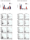
Figure 2. Circulating CD3+ T-cell profiles are similar in atopic dermatitis (AD) and psoriasis
(A) FACS gating used in the analysis of peripheral blood. (B) Frequencies of circulating cytokine-producing CD4+ and CD8+ T-cells did not differ between AD and psoriasis. Graphs summarize cytokine-producing CD4+ and CD8+ T-cell subsets in AD and psoriasis blood. Representative FACS plots are shown.
Figure 3
Figure 3. Atopic dermatitis (AD) and psoriasis lesions have distinct T-cell profiles
(A) Graph of CD4+ T-cell subsets in AD and psoriasis lesions showing Th1/Th17(IFNγ/IL-17) polarization in psoriasis, Th2(IL-4) in AD, and similar IL-22+CD4+ frequencies in both. Representative FACS plots follow. (B) Graph of skin CD8+T-cell subsets showing Tc1(IFNγ) polarity in psoriasis and IL-22+CD8+ polarity in AD. Representative FACS plots follow. *p<0.05, **p<0.01, ***p<0.005.
Figure 4
Figure 4. Unique IL-22-producing CD4+ and CD8+ subsets are abundant in AD lesions
(A) FACS gating used in the analysis of IL-22-producing T-cells. (B) Majority of IL-22+CD4+T-cells in AD are not Th1, Th2 or Th17 cells. Psoriasis IL-22 is produced by Th1, Th17 and “Th22” cells. Representative FACS plots shown. (C) Majority of IL-22-producing CD8+T-cells in AD are “Tc22” cells.
Figure 5
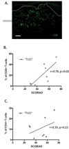
Figure 5. IL-22+CD8+ T-cell frequency correlates with disease severity
(A) Immunofluorescence showing abundant CD8+T-cells in AD dermis. Bar: 100μm. (B) Linear regression showing positive correlation between IL-22-producing CD8+T-cells (“Tc22”) and AD clinical severity (SCORAD) (r=0.78, p<0.05). (C) Linear regression of IL-22-producing CD4+T-cells (“Th22”) showing positive trend with disease severity but not statistically significant.
Figure 6
Figure 6. Model of major T-cells and their cytokines in chronic skin lesions of psoriasis vs. atopic dermatitis (AD)
In psoriasis, Th1 cells produce IFNg, Th17 cells produce both IL-17 and IL-22 cytokines, and “Th22” cells produce IL-22 but not IL-17. Upregulated IL-17 axis in psoriasis results in increased production of anti-microbial proteins, neutrophil chemoattractants and CCL20 chemokine, while upregulated IL-22 causes epidermal acanthosis. In AD, the Th2 cytokines IL-4 and IL-13 potentially inhibit Th17 T-cell activity resulting in reduced IL-17 effects. Both “Th22” and “Tc22” T-cells in AD are responsible for significantly increased IL-22 levels. The upregulated IL-22 in chronic AD skin may explain the marked acanthosis that is histologically similar to psoriasis, and the defective terminal differentiation seen in this disease.

References

    1. Li J, Chen X, Liu Z, Yue Q, Liu H. Expression of Th17 cytokines in skin lesions of patients with psoriasis. J Huazhong Univ Sci Technolog Med Sci. 2007;27:330–2. - PubMed
    1. Lowes MA, Kikuchi T, Fuentes-Duculan J, Cardinale I, Zaba LC, Haider AS, et al. Psoriasis vulgaris lesions contain discrete populations of Th1 and Th17 T cells. J Invest Dermatol. 2008;128:1207–11. - PubMed
    1. Nograles KE, Zaba LC, Guttman-Yassky E, Fuentes-Duculan J, Suarez-Farinas M, Cardinale I, et al. Th17 cytokines interleukin (IL)-17 and IL-22 modulate distinct inflammatory and keratinocyte-response pathways. Br J Dermatol. 2008 - PMC - PubMed
    1. Kryczek I, Bruce AT, Gudjonsson JE, Johnston A, Aphale A, Vatan L, et al. Induction of IL-17+ T cell trafficking and development by IFN-gamma: mechanism and pathological relevance in psoriasis. J Immunol. 2008;181:4733–41. - PMC - PubMed
    1. Zheng Y, Danilenko DM, Valdez P, Kasman I, Eastham-Anderson J, Wu J, et al. Interleukin-22, a T(H)17 cytokine, mediates IL-23-induced dermal inflammation and acanthosis. Nature. 2007;445:648–51. - PubMed

Publication types

MeSH terms