. 2009 Jun 15;180(2):261-72.
doi: 10.1016/j.jneumeth.2009.03.017. Epub 2009 Mar 31.

A white matter stroke model in the mouse: axonal damage, progenitor responses and MRI correlates

Affiliations

A white matter stroke model in the mouse: axonal damage, progenitor responses and MRI correlates

Elif G Sozmen et al. J Neurosci Methods. .

Abstract

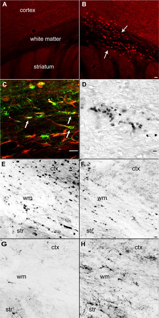
Subcortical white matter stroke is a common stroke subtype but has had limited pre-clinical modeling. Recapitulating this disease process in mice has been impeded by the relative inaccessibility of the subcortical white matter arterial supply to induce white matter ischemia in isolation. In this report, we detail a subcortical white matter stroke model developed in the mouse and its characterization with a comprehensive set of MRI, immunohistochemical, neuronal tract tracing and electron microscopic studies. Focal injection of the vasoconstrictor endothelin-1 into the subcortical white matter produces an infarct core that develops a maximal MRI signal by day 2, which is comparable in relative size and location to human subcortical stroke. Immunohistochemical studies indicate that oligodendrocyte apoptosis is maximal at day 1 and apoptotic cells extend away from the stroke core into the peri-infarct white matter. The amount of myelin loss exceeds axonal fiber loss in this peri-infarct region. Activation of microglia/macrophages takes place at 1 day after injection near injured axons. Neuronal tract tracing demonstrates that subcortical white matter stroke disconnects a large region of bilateral sensorimotor cortex. There is a robust glial response after stroke by BrdU pulse-labeling, and oligodendrocyte precursor cells are initiated to proliferate and differentiate within the first week of injury. These results demonstrate the utility of the endothelin-1 mediated subcortical stroke in the mouse to study post-stroke repair mechanisms, as the infarct core extends through the partially damaged peri-infarct white matter and induces an early glial progenitor response.


Conflict of interest statement

Conflict of interest

The authors declare no conflict of interest.

Figures

Fig. 1
Fig. 1
Human and mouse subcortical white matter stroke. (A) Human FLAIR sequence MRI taken 2 days after left hemispheric subcortical stroke. Arrow denotes white matter hyperintensity that was new in comparison to previous scans from this patient. Case is taken from clinical service of the authors (STC). (B) Mouse MRI taken 2 days after ET-1 injection. Arrow denotes hyperintensity caused by stroke. (C) Nissl staining of subcortical white matter 7 days after stroke. Note small cavity in white matter and adjacent increase in cellularity. (D) White matter stroke in YFP-H mouse line. Left panel is contralateral and white matter is ipsilateral to the stroke 7 days after ET-1 injection. Arrow denotes infarct core, with loss of axons. Note bright axon retraction bulbs in fibers adjacent to the lesion. Bar in (C) = 350 μm. Bar in (D) = 60 μm.
Fig. 2
Fig. 2
Cell death in white matter one day after stroke. (A) Immunohistochemical staining for AIF in contralateral hemisphere. (B) AIF stain in the hemisphere ipsilateral to stroke. Note positive cells in white matter and deepest cortical layers, and linear rows of AIF immunoreactive cells (arrows). (C) CAII stain for oligodendrocytes (green) activated PARP immunoreactivity (red). Note multiple CAII and activated PARP double positive cells in subcortical white matter (D) In situ nick translation showing double stranded DNA nicking in apoptotic cells in subcortical white matter. (E) Transferrin immunoreactive oligodendrocytes in control subcortical white matter region. (F) Transferrin immunoreactive oligodendrocytes 7 days after subcortical white matter stroke. (G) NG2 immunoreactive OPCs in control subcortical white matter region. (H) NG2 immunoreactive OPCs 14 days after subcortical white matter stroke. ctx = cortex, str = striatum, wm = subcortical white matter. Bar in (B) = 50 μm and applies to (A and B); bar in (Cand D) = 40 μm, Bar in (F) = 20 μm and applies to (E–H) (for interpretation of the references to color in this figure legend, the reader is referred to the web version of the article).
Fig. 3
Fig. 3
Glial and inflammatory responses to white matter stroke. (A) GFAP immunoreactivity in subcortical white matter on control brains. (B) GFAP immunoreactivity in subcortical white matter 7 days after stroke. Hyaluronic acid labeling with HABP in subcortical white matter (C) in control brain and (D) in peri-infarct area 7 days after stroke. (E) SMI-32 immunoreactive axons (red) and IBA-1 immunoreactive microglia/macrophages (green) in contralateral subcortical white matter. (F) SMI-32 positive fibers (red) and IBA-1 positive microglia/macrophages (green) 7 days after stroke. Inset shows a high power view of IBA-1 positive activated microglia. Bar in (B) = 40 μm and applies to (A and B); Bar in (D) = 40 μm and applies to (C and D); Bar in (F) = 40 μm and applies to (E and F) (for interpretation of the references to color in this figure legend, the reader is referred to the web version of the article).
Fig. 4
Fig. 4
Axonal and myelin damage after subcortical stroke. (A and B) MBP immunoreactivity contralateral (A) and ipsilateral (B) to stroke at 28 days after the infarct. Note area of stroke seen as a loss of myelin in a hole in subcortical white matter (arrows). (C and D) Neurofilament immunoreactivity contralateral (C) and ipsilateral (D) to stroke in same section as (A and B). The stroke site is apparent as an area of neurofilament/axonal loss (arrows). (E and F) MBP and SMI-31 stain of myelin and neurofilament patterns in contralateral (E) and ipsilateral (F) subcortical white matter 7 days after stroke. Arrows in (F) highlight the area of diminished MBP staining and preserved axonal filament staining. (G and H) High power views of peri-infarct white matter 7 days after stroke. In contralateral white matter (E) axons (red) are closely associated with myelin staining (green). At the stroke site axonal profiles (red) are present in areas of diminished myelin staining. Bar in (B) = 40 μm and applies to (A–F). Bar in (H) = 40 μm and applies to (G and H) (for interpretation of the references to color in this figure legend, the reader is referred to the web version of the article).
Fig. 5
Fig. 5
Axonal damage from ischemic white matter stroke. (A–D) Sections taken from YFP-H line 7 days after stroke. (A) Normal subcortical white matter contains multiple linear axons and axon fascicles. (B) Seven days after white matter stroke axons have dropped out leading to fewer labeled axons in subcortical white matter. Damaged axons contain retraction bulbs seen as bright varicosities. (C) Higher power view of damaged axons and retraction bulbs present in white matter projections through the striatum below the stroke site. (D) Higher power view of retraction bulbs and beaded axons (arrows) in subcortical white matter. (E and F) Electron micrograph of subcortical white matter 1 day after stroke (E) and 7 days after stroke (F). Arrows in (E) show injured axons with electron dense cytoplasm. Arrows in (F) show degenerating axons with vacuoles and separated and fragmented myelin sheaths. Arrowhead in (F) shows cell with microglial morphology adjacent to degenerating axon EM images taken at 4800×. Bar in (B) applies to (A and B) = 40 μm; Bar in (D) applies to (C and D) = 40 μm.
Fig. 6
Fig. 6
Neuronal projections through stroke site. Line drawings are from digitized maps of labeled cells from a co-injection of the anatomical tracer BDA with ET-1. The sites of the stroke and BDA injections are indicated with X. The dots represent cells that were retrogradely labeled from the stroke site. Bar = 350 μm. The inset is a high magnification photomicrograph of labeled neurons from the BDA injection. Bar in inset = 20 μm.
Fig. 7
Fig. 7
Early and late response of OPCs and oligodendrocytes following subcortical stroke. BrdU pulse-chase experiment at day 7 and 28 after subcortical stroke. (A) Representative images of BrdU double labeling of oligodendrocyte progenitor cells (OPC) with OPC marker NG2 (top panel), and oligodendrocytes with specific marker transferrin (bottom panel) at day 7. The insets correspond to higher power views. Bar in right middle panel of A = 20 μm. (B) Total number of double labeled cells estimated by stereology: means ± standard deviations. NG2 7 day vs. 28 day 3918.33 ± 238.03, 2150.0 ± 413.54, p = 0.003; transferrin 7 day vs. 28 day 3665.0 ± 456.54, 1941.67 ± 677.98, p = 0.02.

References

    1. Back SA, Tuohy TM, Chen H, Wallingford N, Craig A, Struve J, et al. Hyaluronan accumulates in demyelinated lesions and inhibits oligodendrocyte progenitor maturation. Nat Med. 2005;11:966–72. - PubMed
    1. Bamford J, Sandercock P, Dennis M, Burn J, Warlow C. Classification and natural history of clinically identifiable subtypes of cerebral infarction. Lancet. 1991;337:1521–6. - PubMed
    1. Carmichael ST. Animal models of stroke: size, mechanism and purpose. NeuroRx. 2005;2:396–409. - PMC - PubMed
    1. Carmichael ST, Vespa PM, Saver JL, Coppola G, Geschwind DH, Starkman S, et al. Genomic profiles of damage and protection in human intracerebral hemorrhage. J Cereb Blood Flow Metab. 2008;28:1860–75. - PMC - PubMed
    1. Dimou L, Simon C, Kirchhoff F, Takebayashi H, Götz M. Progeny of Olig2-expressing progenitors in the gray and white matter of the adult mouse cerebral cortex. J Neurosci. 2008;28:10434–42. - PMC - PubMed

Publication types

MeSH terms