. 2009 Jul 31;284(31):20718-28.
doi: 10.1074/jbc.M806711200. Epub 2009 May 13.

Roles of human AND-1 in chromosome transactions in S phase

Affiliations

Roles of human AND-1 in chromosome transactions in S phase

Naoko Yoshizawa-Sugata et al. J Biol Chem. .

Abstract

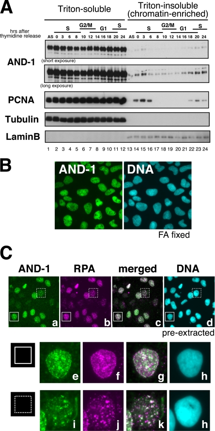
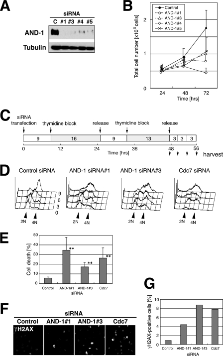
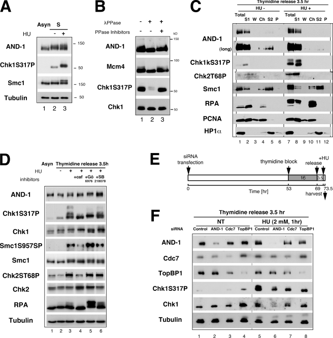
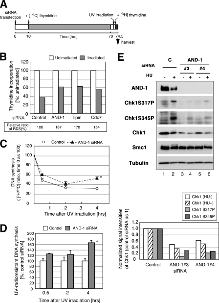
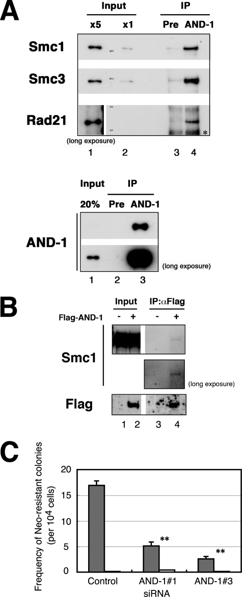
Coordinated execution of DNA replication, checkpoint activation, and postreplicative chromatid cohesion is intimately related to the replication fork machinery. Human AND-1/chromosome transmission fidelity 4 is localized adjacent to replication foci and is required for efficient DNA synthesis. In S phase, AND-1 is phosphorylated in response to replication arrest in a manner dependent on checkpoint kinase, ataxia telangiectasia-mutated, ataxia telangiectasia-mutated and Rad3-related protein, and Cdc7 kinase but not on Chk1. Depletion of AND-1 increases DNA damage, delays progression of S phase, leads to accumulation of late S and/or G2 phase cells, and induces cell death in cancer cells. It also elevated UV-radioresistant DNA synthesis and caused premature recovery of replication after hydroxyurea arrest, indicating that lack of AND-1 compromises checkpoint activation. This may be partly due to the decreased levels of Chk1 protein in AND-1-depleted cells. Furthermore, AND-1 interacts with cohesin proteins Smc1, Smc3, and Rad21/Scc1, consistent with proposed roles of yeast counterparts of AND-1 in sister chromatid cohesion. Depletion of AND-1 leads to significant inhibition of homologous recombination repair of an I-SceI-driven double strand break. Based on these data, we propose that AND-1 coordinates multiple cellular events in S phase and G2 phase, such as DNA replication, checkpoint activation, sister chromatid cohesion, and DNA damage repair, thus playing a pivotal role in maintenance of genome integrity.


Figures

FIGURE 1.
FIGURE 1.
Expression and cellular localization of human AND-1 protein during cell cycle. A, HeLa cells were released from double thymidine block for the indicated time (hours) and fractionated into Triton-soluble and -insoluble fractions. Proteins were analyzed by Western blotting using the antibodies indicated. The affinity-purified anti-AND-1Npep antibody was used. S, S phase; G2/M, G2 phase and mitosis; G1, G1 phase. B, indirect immunofluorescence microscopy. U2OS cells fixed with paraformaldehyde (FA) were stained with anti-AND-1C antibody. DNA was counterstained with DAPI. C, U2OS cells were pre-extracted with detergent and immunostained with anti-AND-1C, anti-RPA, and DAPI (top panels). Enlarged images of two areas (indicated by solid line or dotted line square) in top panels are shown below.
FIGURE 2.
FIGURE 2.
Knockdown of AND-1 delays S phase progression and induces DNA damage and cell death. A, U2OS cells were transfected with four different siRNAs against AND-1 (#1, #3, #4, or #5) or control (C) siRNA for 72 h. Twenty-five-μg proteins of whole cell extracts were analyzed by immunoblot using the antibodies indicated. The affinity-purified anti-AND-1NPep antibody was used. B, numbers of viable HeLa cells transfected with AND-1 siRNAs were counted at 24, 48, or 72 h after transfection. Black circle, control siRNA; open circle, AND-1 siRNA number 1; open triangle, AND-1 siRNA number 3; open diamond, AND-1 siRNA number 4; cross, AND-1 siRNA number 5. Error bars indicate S.D. of three independent transfection experiments. C, time schedule of the experiment in D as described under “Experimental Procedures.” D, DNA contents of siRNA-treated HeLa cells at various time points after double thymidine block and release. E, knockdown of AND-1 induces cell death. HeLa cells were transfected with the indicated siRNA, and the population of dead cells was counted at 77 h as described under “Experimental Procedures.” Error bars indicate S.D. Data with asterisks revealed significant differences compared with control siRNA-transfected cells (p < 0.01). F, DNA damage signals increased by knockdown of AND-1 protein. AND-1-depleted HeLa cells were stained with anti-γH2AX antibody at 72 h after transfection. G, populations of γH2AX-positive cells in F were counted and presented.
FIGURE 3.
FIGURE 3.
Mobility shift of AND-1 after replication stress depends on ATR/ATM and Cdc7 but not on Chk1 kinase. A, whole cell extracts were prepared from asynchronously (Asyn) growing (lane 1) or S phase-synchronized cells (3.5 h release from single thymidine block; lanes 2 and 3). In lane 3, cells were treated with 5 mm HU for 1 h before harvest. Indicated proteins were detected by immunoblot analysis. B, 30 μg of the whole cell extracts from the HU-treated cells (lane 3 of A) was subjected to phosphatase (λPPase) assay as described under “Experimental Procedures.” After treatment, samples were separated on SDS-PAGE, and proteins were detected by immunoblot with the indicated antibodies. C, HeLa cells, synchronized in S phase by release for 3.5 h from double thymidine block, were harvested untreated (HU−) or after additional incubation with 5 mm HU for 2 h (HU+). Cells were sequentially fractionated into Triton-soluble fractions (S1), washed fractions after extraction (W), supernatants after DNase I digestion (Ch), 0.5 m NaCl-soluble fractions (S2), and insoluble fractions (P). S1 fraction (20 μg of protein) and other fractions from equivalent amounts of cells were analyzed by immunoblot using the indicated antibodies. Total, whole cell extracts from the same numbers of the cells. D, effects of kinase inhibitors on AND-1 protein and other replication/checkpoint proteins. Asynchronous or S phase-synchronized (lanes 2–6; 3.5 h release from single thymidine block) HeLa cells were incubated with HU (2 mm) for 1 h. In lanes 4–6, the following reagents were added at 1.5 h before HU treatment; 5 mm caffeine (caf); 1 μm Gö6976; 5 μm SB218078. Twenty μg of whole cell extracts were resolved on SDS-PAGE, and proteins were detected with indicated antibodies. E, time schedule of the experiment in F as described under “Experimental Procedures.” F, HeLa cells were transfected with siRNA as shown, synchronized at S phase by 3.5 h release from thymidine block, and treated with HU. Whole cell extracts (20 μg of protein) were resolved on SDS-PAGE, and proteins were detected with indicated antibodies. Anti-AND-1C (in A, B, D, and F) or affinity-purified anti-AND-1NPep antibody (in C) and AND-1 number 3 siRNA (in F) were used.
FIGURE 4.
FIGURE 4.
UV radio-resistant DNA synthesis (UV-RDS) in AND-1 knockdown U2OS cells. A, time schedule of the experiment. At 10 h after transfection with control or AND-1 number 3 siRNA, HeLa cells were transferred to the medium containing 20 nCi of [14C]thymidine per ml, followed by incubation for 60 h. After incubation with nonisotopic medium for 2 h, cells were irradiated with UVC (5 J/m2, UV) or mock-irradiated. After 1 h, 10 μCi of [3H]thymidine was added per ml of medium, and incubation was continued for 1.5 h. Cells were harvested, and incorporated radioactivity was measured by scintillation counter. B, ratio of 3H/14C incorporation of AND-1, Tipin, and Cdc7-depleted cells. The values of the unirradiated sample were taken as 100 in each siRNA-treated cells. At the bottom, RDS is shown as values relative to control siRNA, set as 100. We repeated the experiments three times using U2OS or HeLa cells, and the representative data are shown here. C and D, RDS was assessed in time course using the similar protocol described in A except for intensity of UV irradiation (3 J/m2) and for incorporation time of [3H]thymidine (30 min before indicated time). Ratio of 3H/14C incorporation at each time point was plotted. The efficiency of DNA synthesis of unirradiated cells (in C) or that of control siRNA-treated cells after irradiation (in D) was set as 100. Data with asterisks showed significant differences compared with control cells (p < 0.05). Open circles, control siRNA; filled triangles, AND-1 number 4 siRNA. E, knockdown of AND-1 leads to reduction of Chk1 protein level. HeLa cells were transfected with control (C) or AND-1 number 3 or number 4 siRNA and incubated with 2 mm HU for 1 h (+) or untreated (−) before harvest at 48 h after transfection. Twenty μg of protein was resolved on SDS-PAGE, and proteins were detected by the antibodies indicated. The anti-AND-1C antibody was used. In the bottom panel, the signal intensities of Chk1 (untreated, open column; HU-treated, shaded), Chk1 S317P (HU-treated, gray), and Chk1 S345P (HU-treated, dark gray) were normalized with those of α-tubulin. We repeated the experiments five times, and representative data are shown.
FIGURE 5.
FIGURE 5.
AND-1 interacts with cohesin complex and is required for recombinational repair. A, nuclear extracts (700 μg of protein in top panel; 30 μg of proteins in bottom panel) of HeLa S3 cells were immunoprecipitated (IP) with anti-AND-1C antibody or preimmune serum (Pre) and subjected to immunoblot analysis. In the top panel, input (x5 in lane 1 and x1 in lane 2) represents 0.1 or 0.02%, respectively, of the starting extracts used for immunoprecipitation in lanes 3 and 4. In the bottom panel, input represents 20% of the starting extracts used in lane 3. The asterisk indicates a nonspecific signal. B, HeLa cells were transfected with pME18S-FLAG-AND-1 (+) (lanes 2 and 4) or pME18S (−) (lanes 1 and 3) and harvested at 48 h after transfection. Cells were lysed and immunoprecipitated (IP) with anti-FLAG M2-agarose and analyzed for co-immunoprecipitation of Smc1 protein. Input (lanes 1 and 2) represents 10 μg of the starting extracts used for immunoprecipitation in lanes 3 and 4, respectively. In B, contrast-enhanced images of long exposed films are also shown. C, analysis of I-SceI-induced homologous recombinational repair using a neomycin-based reporter construct. The frequency of neomycin-resistant colonies was counted in control, AND-1 number 1, or AND-1 number 3 siRNA-transfected SW480sn3 cells. The error bars represent S.D. from three independent experiments, and p values are less than 0.01 for both AND-1 number 1 and AND-1 number 3 siRNAs compared with control. Values of cells transfected with I-SceI pCMV3his-I-SceI (gray bars) or empty vector (open bars) are shown. Similar defects in recombinational repair were observed also in cells transfected with AND-1 number 4 and number 5 siRNA (data not shown).

Similar articles

Cited by

References

    1. Zou L., Stillman B. (1998) Science 280, 593–596 - PubMed
    1. Mimura S., Takisawa H. (1998) EMBO J. 17, 5699–5707 - PMC - PubMed
    1. Masai H., Taniyama C., Ogino K., Matsui E., Kakusho N., Matsumoto S., Kim J. M., Ishii A., Tanaka T., Kobayashi T., Tamai K., Ohtani K., Arai K. (2006) J. Biol. Chem. 281, 39249–39261 - PubMed
    1. Takayama Y., Kamimura Y., Okawa M., Muramatsu S., Sugino A., Araki H. (2003) Genes Dev. 17, 1153–1165 - PMC - PubMed
    1. Tanaka S., Umemori T., Hirai K., Muramatsu S., Kamimura Y., Araki H. (2007) Nature 445, 328–332 - PubMed

Publication types

MeSH terms