. 2009 Jul;20(13):3064-76.
doi: 10.1091/mbc.e08-12-1223. Epub 2009 May 13.

Rec8 guides canonical Spo11 distribution along yeast meiotic chromosomes

Affiliations

Rec8 guides canonical Spo11 distribution along yeast meiotic chromosomes

Kazuto Kugou et al. Mol Biol Cell. 2009 Jul.

Abstract

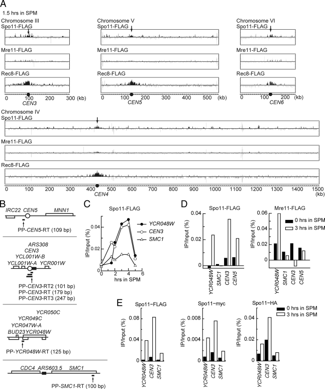
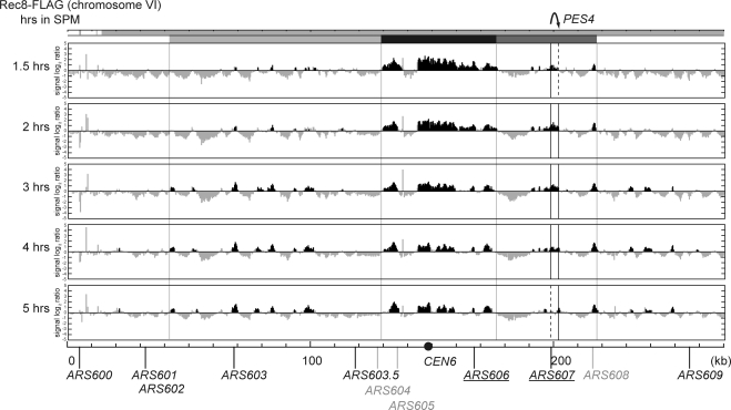
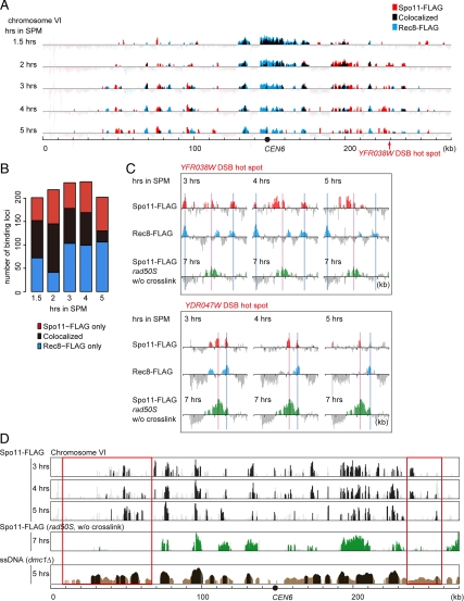
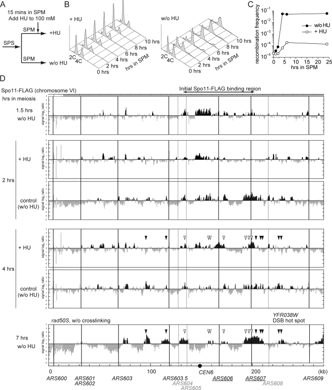
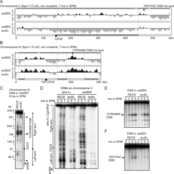
Spo11-mediated DNA double-strand breaks (DSBs) that initiate meiotic recombination are temporally and spatially controlled. The meiotic cohesin Rec8 has been implicated in regulating DSB formation, but little is known about the features of their interplay. To elucidate this point, we investigated the genome-wide localization of Spo11 in budding yeast during early meiosis by chromatin immunoprecipitation using high-density tiling arrays. We found that Spo11 is dynamically localized to meiotic chromosomes. Spo11 initially accumulated around centromeres and thereafter localized to arm regions as premeiotic S phase proceeded. During this stage, a substantial proportion of Spo11 bound to Rec8 binding sites. Eventually, some of Spo11 further bound to both DSB and Rec8 sites. We also showed that such a change in a distribution of Spo11 is affected by hydroxyurea treatment. Interestingly, deletion of REC8 influences the localization of Spo11 to centromeres and in some of the intervals of the chromosomal arms. Thus, we observed a lack of DSB formation in a region-specific manner. These observations suggest that Rec8 would prearrange the distribution of Spo11 along chromosomes and will provide clues to understanding temporal and spatial regulation of DSB formation.


Figures

Figure 1.
Figure 1.
Distributions of Spo11-FLAG and Mre11-FLAG on meiotic chromosome VI. (A) Summary of meiotic events in SK1 background strain. Rep, premeiotic DNA replication; DSB, DSB formation; Rec, homologous recombination; MI, meiosis I. (B and C) ChIP-chip analyses of Spo11-FLAG (B) and Mre11-FLAG (C). RKD1311 (SPO11-FLAG) and RKD1313 (MRE11-FLAG) cells were cross-linked at indicated time points after the medium change and analyzed. The horizontal axis represents the physical position on the chromosome. The vertical axis represents the relative binding strength of these proteins as compared with the Input signals. The scale of the vertical axis is expressed in log2. Black and light gray vertical bars represent detection loci showing significant binding ratios and insufficient enrichment in the immunoprecipitated fraction, respectively. The position of the centromere is indicated with the black circle on the horizontal axis. Timing of the Spo11-FLAG and Mre11-FLAG binding along chromosome VI is indicated by shaded boxes (from black to white) above the top panel. Shaded parts under the coordinate axis in B represent detection loci for the mapping (for details, see Supplemental Figure S1). Names and locations of known ARSs are shown at the bottom of each figure. Underlined ARS and light gray-colored ARS represent active early and inactive replication origins, respectively.
Figure 2.
Figure 2.
Spo11-FLAG localized to the centromeres at early meiosis. (A) Localization of Spo11-FLAG (RKD1311), Mre11-FLAG (RKD1313), and Rec8-FLAG (RKD1317) at 1.5 h after transfer to SPM on chromosome III–VI. All indications are as described in Figure 1. (B) Map of the primer pairs (PP) used for quantitative real-time PCR (qPCR). Open circle, open boxes, closed boxes, and horizontal thick bars indicate centromere, open reading frames, ARSs and PCR fragments, respectively. (C) ChIP-qPCR of Spo11-FLAG. The Spo11-FLAG binding to CEN3 was analyzed by qPCR using PP-CEN3-RT. (D) Localization of Spo11-FLAG and Mre11-FLAG at centromeres, a DSB hot spot and a DSB cold spot. The bindings of Spo11-FLAG and Mre11-FLAG to CEN3 and CEN5 were detected by qPCR using primer pairs PP-CEN5-RT and PP-CEN3-RT2, respectively. The ratio (immunoprecipitated/input) was calculated by subtracting the value of no-tagged sample from that of FLAG-tagged sample. (E) Localization of Spo11 to CEN3 was not affected by different epitope tags. FLAG-tagged (RKD1311), HA-tagged (RKD1329), and myc-tagged Spo11 (RKD1339) were detected on CEN3 at 3 h in SPM. PP-CEN3-RT3 was used for qPCR.
Figure 3.
Figure 3.
Distributions of Rec8-FLAG on meiotic chromosome VI. All indications are as described in Figure 1. RKD1317 (REC8-FLAG) was used in this analysis.
Figure 4.
Figure 4.
Comparison of DSB sites and binding sites of Spo11-FLAG and Rec8-FLAG. (A) Overlay of Spo11-binding sites (red bars) with Rec8-binding sites (blue bars) on chromosome VI. Overlapped sites are shown as black bars. Other indications are as described in Figure 1. Only detection loci judged as significant binding are shown. (B) Stacked bar graph of numbers of binding loci for Spo11-FLAG only (red), Rec8-FLAG only (blue), and both proteins (black). (C) Magnified views of localization of Spo11-FLAG, Rec8-FLAG, and DSBs (rad50S) around YFR038W (top) and YDR047W DSB (bottom) hot spots during 3–5 h after meiotic induction. The DSB sites were revealed as Spo11-FLAG binding sites by ChIP-chip analysis without cross-linking in RKD1325 (rad50S SPO11-FLAG) at 7 h after meiotic induction. Spo11-FLAG binding sites, Rec8-FLAG binding sites, and DSB sites (rad50S) are indicated with vertical red, blue, and green bars, respectively. Light red and blue vertical lines represent positions of DSB sites and Rec8-FLAG binding sites, respectively. (D) Comparison of binding sites of Spo11-FLAG in wild-type and DSB sites in rad50S (green bars) and dmc1Δ (brown bars). Mapping of the DSB sites in dmc1Δ was carried out according to Buhler's microarray data (Buhler et al., 2007; GEO accession GSE8981). The signal (enrichment of single-stranded DNA) is transformed to log2. In the lowest panel (single-stranded [ss]DNA dmc1Δ), light and dark brown bars represent weak DSB sites (enrichment >2) and strong DSB site (enrichment >5), respectively. DSB cold domains observed only in rad50S are indicated with red boxes.
Figure 5.
Figure 5.
Effects of blocking replication by HU treatment on the distributions of Spo11-FLAG. (A) The culture (RKD1311) was divided into two parts on transferring to SPM. HU was added to one of them 15 min after meiosis induction. (B) Progression of premeiotic S phase monitored by FACS analyses. (C) Recombination frequency measured by return-to-growth at the arg4-bgl/arg4-nsp heteroallele. (D) Effects of 100 mM HU treatment on the association of Spo11-FLAG with meiotic chromosome VI. Cells from the same culture as in (B) were taken at 2 and 4 h of meiosis and analyzed. The binding sites of Spo11-FLAG were classified into two subgroups as indicated with filled and open arrowheads (see text). Other indications are as described in Figure 1.
Figure 6.
Figure 6.
Effect of REC8 deletion on the distribution of Spo11-FLAG. (A) Distribution of Spo11-FLAG on chromosome III in rec8Δ cells (RKD1319, left) and REC8+ cells (RKD1311, right). Chromosome domains harboring the Spo11-FLAG binding sites in rec8Δ are indicated with light gray boxes on the horizontal axis. All other indications are as described in Figure 1. (B) Distribution of Spo11-FLAG on chromosome V in rec8Δ cells (top) and REC8+ cells (bottom). The position of CEN5 is indicated with arrows and closed circles.
Figure 7.
Figure 7.
Effect of REC8 deletion on the distribution of DSB sites. (A and B) DSB sites on chromosome V (A) and chromosome III (B) determined by ChIP-chip analyses using RKD1327 (rec8Δ rad50S SPO11-FLAG) and RKD1325 (rad50S SPO11-FLAG). (C) DSB analysis using pulsed-field gel electrophoresis followed by Southern blotting with a chromosome III left-end probe (shown in closed box). RKD1321 (rad50S) and RKD1323 (rad50S rec8Δ) were used. Closed circle indicates the centromere. (D) DSB analysis using pulsed-field gel electrophoresis followed by Southern blotting using a probe for a sequence of the chromosome V left-arm (shown in closed box). YKT23 (dmc1Δ), YKT28 (dmc1Δ rec8Δ), RKD1321 (rad50S), and RKD1323 (rad50S rec8Δ) were used. Closed circle indicates the centromere. (E and F) DSBs around the YCR048W (E) and YER153C (F) DSB hot spots. The DSBs were detected by Southern blotting of genomic DNA digested with BglII (YCR048W) or HindIII (YER153C). Note that the YCR048W DSB sites (open arrowhead) are active also rec8Δ, but the YER153C DSB site (closed arrowhead) is not.

References

    1. Alani E., Padmore R., Kleckner N. Analysis of wild-type and rad50 mutants of yeast suggests an intimate relationship between meiotic chromosome synapsis and recombination. Cell. 1990;61:419–436. - PubMed
    1. Baudat F., Nicolas A. Clustering of meiotic double-strand breaks on yeast chromosome III. Proc. Natl. Acad. Sci. USA. 1997;94:5213–5218. - PMC - PubMed
    1. Bishop D. K., Park D., Xu L., Kleckner N. DMC1: a meiosis-specific yeast homolog of E. coli recA required for recombination, synaptonemal complex formation, and cell cycle progression. Cell. 1992;69:439–456. - PubMed
    1. Blat Y., Protacio R. U., Hunter N., Kleckner N. Physical and functional interactions among basic chromosome organizational features govern early steps of meiotic chiasma formation. Cell. 2002;111:791–802. - PubMed
    1. Blitzblau H. G., Bell G. W., Rodriguez J., Bell S. P., Hochwagen A. Mapping of meiotic single-stranded DNA reveals double-stranded-break hotspots near centromeres and telomeres. Curr. Biol. 2007;17:2003–2012. - PubMed

Publication types

MeSH terms