Comparative Study
. 2009 Aug;83(15):7749-60.
doi: 10.1128/JVI.00108-09. Epub 2009 May 13.

Features distinguishing Epstein-Barr virus infections of epithelial cells and B cells: viral genome expression, genome maintenance, and genome amplification

Affiliations
Comparative Study

Features distinguishing Epstein-Barr virus infections of epithelial cells and B cells: viral genome expression, genome maintenance, and genome amplification

Claire Shannon-Lowe et al. J Virol. 2009 Aug.

Abstract

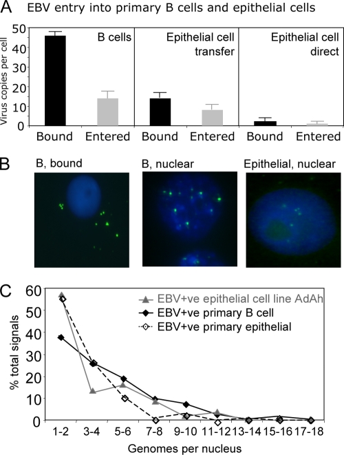
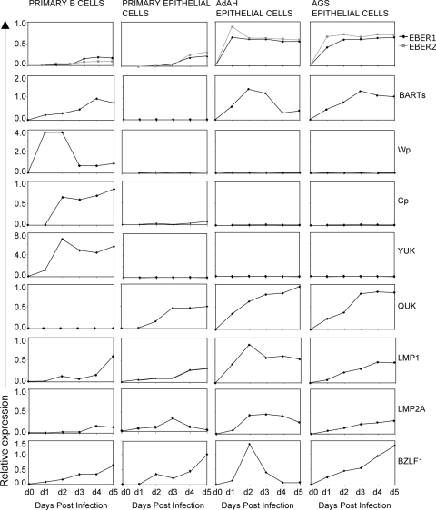
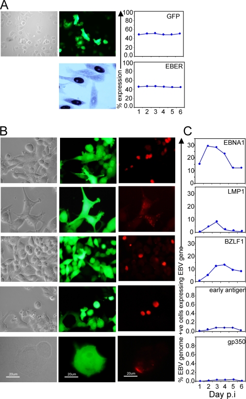
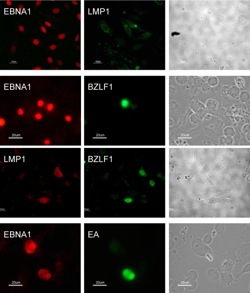
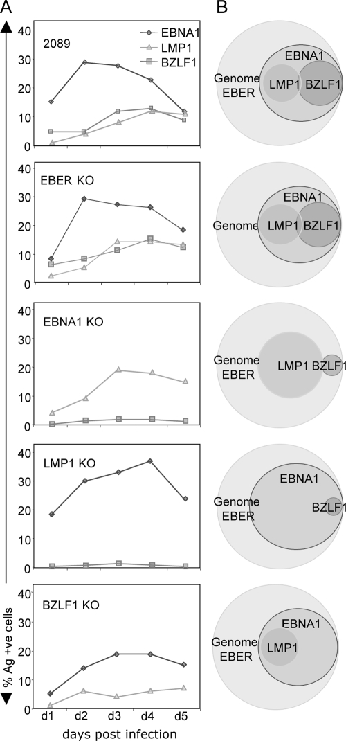
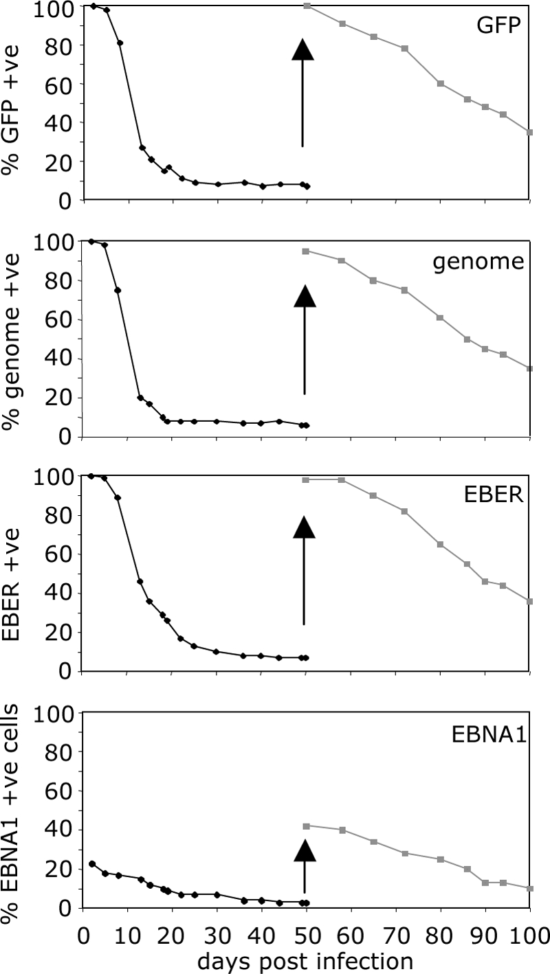
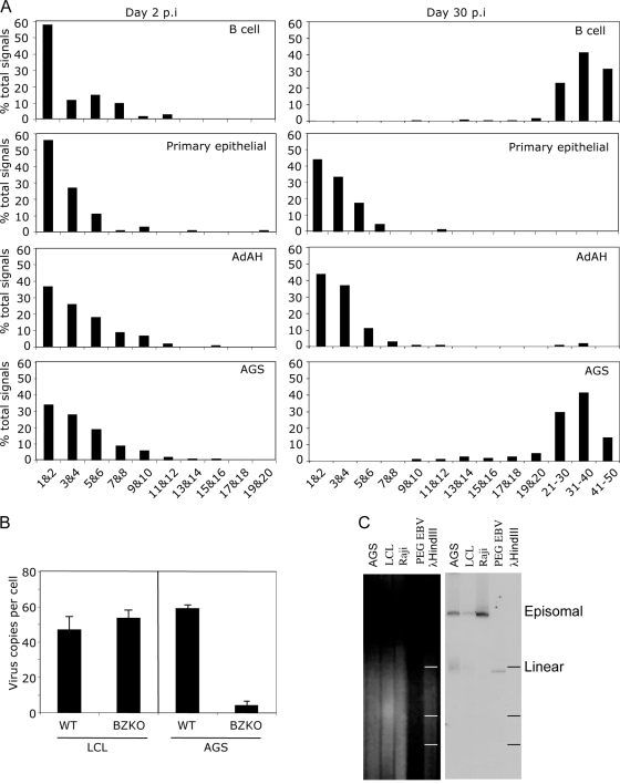
Epstein-Barr virus (EBV) is associated with malignant diseases of lymphoid and epithelial cell origin. The tropism of EBV is due to B-cell-restricted expression of CD21, the major receptor molecule for the virus. However, efficient infection of CD21- epithelial cells can be achieved via transfer from EBV-coated B cells. We compare and contrast here the early events following in vitro infection of primary B cells and epithelial cells. Using sensitive, quantitative reverse transcription-PCR assays for several latent and lytic transcripts and two-color immunofluorescence staining to analyze expression at the single cell level, we confirmed and extended previous reports indicating that the two cell types support different patterns of transcription. Furthermore, whereas infection of B cells with one or two copies of EBV resulted in rapid amplification of the viral genome to >20 copies per cell, such amplification was not normally observed after infection of primary epithelial cells or undifferentiated epithelial lines. In epithelial cells, EBNA1 expression was detected in only ca. 40% of EBER+ cells, and the EBV genome was subsequently lost during prolonged culture. One exception was that infection of AGS, a gastric carcinoma line, resulted in maintenance of EBNA1 expression and amplification of the EBV episome. In contrast to B cells, where amplification of the EBV episome occurred even with a replication-defective BZLF1-knockout virus, amplification in AGS cells was dependent upon early lytic cycle gene expression. These data highlight the influence of the host cell on the outcome of EBV infection with regard to genome expression, amplification, and maintenance.


Figures

FIG. 1.
FIG. 1.
EBV entry into B cells and primary epithelial cells. Virus binding and internalization assays on primary B cells and on epithelial cells infected by transfer infection and direct infection using purified infectious EBV particles at an MOI of 50. (A) Q-PCR assays were used to measure the amount of virus bound to the cell surface and the amount of virus that had entered after incubation at 37°C for 24 h. The results are the means ± the standard deviations of six independent B-cell infection experiments and four independent primary epithelial cell experiments. (B) Detection of viral genomes by FISH. Primary B cells and primary epithelial cells were exposed to purified EBV as described above, and extracellular or internalized genomes were detected by green fluorescence. Nuclei are identified by DAPI staining. (C) Distribution of EBV genome numbers detectable per primary B-cell, primary epithelial cell, and AdAH epithelial cell line. The results are expressed as the percentage of cells with 1 to 18 genomes per nucleus.
FIG. 2.
FIG. 2.
Kinetics of viral transcription after infection of B cells and epithelial cells. EBV transcripts were assayed by quantitative RT-PCR after infection of primary B cells, primary epithelial cells, and the AdAH epithelial cell line. The results for BARTs, EBERs, Wp, Y-U-K EBNA1, LMP1, and LMP2 transcripts are displayed as gene expression relative to a latency III, Wp-using reference LCL, X50-7; Cp transcripts are displayed relative to a latency III, Cp-using reference LCL, Oku-LCL; Q-U-K EBNA1 transcripts are expressed relative to a reference latency I Burkitt's lymphoma line, Rael; and BZLF1 transcripts are expressed relative to a reference LCL, Chege-LCL, containing ca. 2% cells in lytic cycle.
FIG. 3.
FIG. 3.
Viral gene expression at the single epithelial cell level. (A) Photomicrographs of phase-contrast, GFP fluorescence (upper panels) and EBER ISH (lower panel) of AdAH cultures at 72 h postinfection. GFP and EBER expression were quantified and are represented as the percent expression for an average experiment up to 6 days postinfection. (B) Photomicrographs of phase-contrast and GFP fluorescence of AdAH cells sorted for GFP expression 24 h postinfection. Cells were stained for expression of the latent gene products EBNA1 and LMP1 plus the lytic gene products BZLF1, early antigen EA, and the late antigen gp350. (C) Expression of the latent and lytic gene products was quantitated daily to 6 days postinfection from a population of 100% infected (GFP +ve) cells.
FIG. 4.
FIG. 4.
Heterogeneous viral gene expression at the single-cell level. Phase-contrast and fluorescence images of EBV gene expression show heterogeneous gene expression in different subpopulations of AdAH epithelial cells: EBNA1 alone (latency I); EBNA1 and LMP1 (latency II); and EBNA1, BZLF1, ±LMP1, ±EA (abortive lytic).
FIG. 5.
FIG. 5.
Recombinant EBV gene knockout infected epithelial cells. AdAH epithelial cells were infected with either wild-type, EBER1/2 KO, EBNA1 KO, LMP1 KO, or BZLF1 KO recombinant viruses. The cells were sorted for GFP expression at 18 h postinfection, and the cells were analyzed daily for the viral genome (FISH); for expression of EBER1/2 (ISH); and for expression of EBNA1, LMP1, and BZLF1 (immunofluorescence). The results are displayed as the percent expression of EBNA1, LMP1, and BZLF1 in 100% infected cells (GFP +ve) over 5 days postinfection (A) and as Venn diagrams to identify the subpopulations of cells expressing one or more EBV genes (B).
FIG. 6.
FIG. 6.
Maintenance of the viral genome in epithelial cells. AdAH epithelial cells were infected with wild-type virus, sorted for GFP expression at 24 h postinfection, and assayed at every passage over 50 days for GFP expression, viral genome (FISH), EBER1/2 expression (ISH), and EBNA1 expression (immunofluorescence). The cells were then resorted for GFP expression at day 50 and assayed for a further 50 days for the above. The results are displayed as the percent expression of each parameter over 100 days.
FIG. 7.
FIG. 7.
Amplification of the viral genome. (A) Primary B cells and primary epithelial cells (upper two panels), AdAH epithelial cells (third panels), and AGS epithelial cells (fourth panels) were infected with wild-type recombinant EBV and sorted for GFP expression at 48 h postinfection. The infected cells were examined immediately or after 30 days for viral genome copy number per cell by FISH. The results are displayed as the numbers of viral genomes per cell at 48 h postinfection (left-hand panels) and at 30 days postinfection (right-hand panels). (B) Detection of viral genomes by FISH. Primary B cells and AGS cells were infected at an MOI of 1 with wild-type or BZLF1 KO viruses. The cells were examined at 40 days postinfection for genome amplification. Viral genomes are detected by green fluorescence, and nuclei are identified by DAPI staining. (C) Detection of circular and linear viral genomes by Gardella gel analysis and Southern blotting. PEG-precipitated viral particles were used as a control for linear viral genomes, Raji cells were used as a control for episomal DNA, and LCLs were used as a control for both episomal and linear DNA. The vast majority of viral DNA in AGS cells with amplified viral genomes corresponds to episomal DNA, with a very small proportion being linear.

References

    1. Babcock, G. J., L. L. Decker, M. Volk, and D. A. Thorley-Lawson. 1998. EBV persistence in memory B cells in vivo. Immunity 9395-404. - PubMed
    1. Becker, J., U. Leser, M. Marschall, A. Langford, W. Jilg, H. Gelderblom, P. Reichart, and H. Wolf. 1991. Expression of proteins encoded by Epstein-Barr virus transactivator genes depends on the differentiation of epithelial cells in oral hairy leukoplakia. Proc. Natl. Acad. Sci. USA 888332-8336. - PMC - PubMed
    1. Bell, A. I., K. Groves, G. L. Kelly, D. Croom-Carter, E. Hui, A. T. Chan, and A. B. Rickinson. 2006. Analysis of Epstein-Barr virus latent gene expression in endemic Burkitt's lymphoma and nasopharyngeal carcinoma tumour cells by using quantitative real-time PCR assays. J. Gen. Virol. 872885-2890. - PubMed
    1. Brooks, L., Q. Y. Yao, A. B. Rickinson, and L. S. Young. 1992. Epstein-Barr virus latent gene transcription in nasopharyngeal carcinoma cells: coexpression of EBNA1, LMP1, and LMP2 transcripts. J. Virol. 662689-2697. - PMC - PubMed
    1. Busson, P., Q. Zhang, J. M. Guillon, C. D. Gregory, L. S. Young, B. Clausse, M. Lipinski, A. B. Rickinson, and T. Tursz. 1992. Elevated expression of ICAM1 (CD54) and minimal expression of LFA3 (CD58) in Epstein-Barr-virus-positive nasopharyngeal carcinoma cells. Int. J. Cancer 50863-867. - PubMed

Publication types

LinkOut - more resources