. 2009 May 13;29(19):6105-13.
doi: 10.1523/JNEUROSCI.0704-09.2009.

Increased dependence of action selection on recent motor history in Parkinson's disease

Affiliations

Increased dependence of action selection on recent motor history in Parkinson's disease

Rick C Helmich et al. J Neurosci. .

Abstract

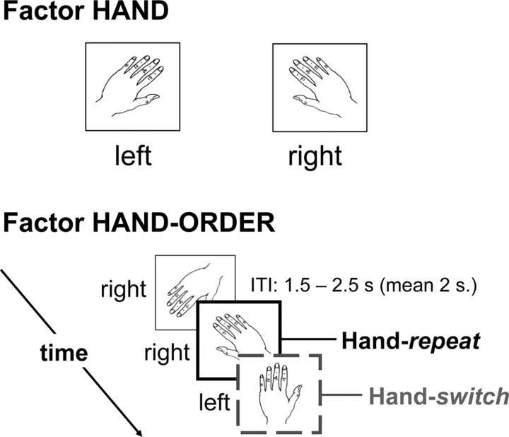
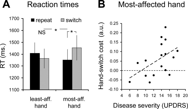
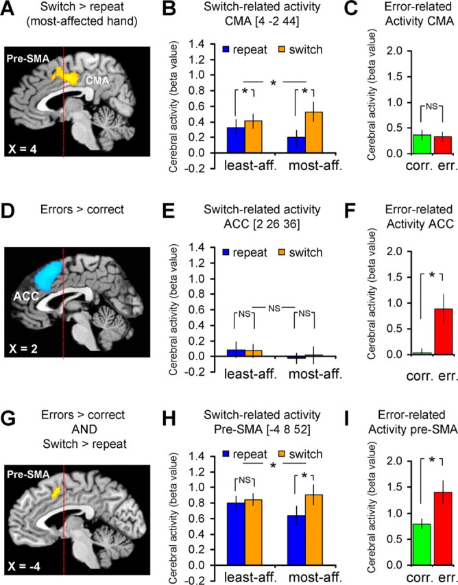
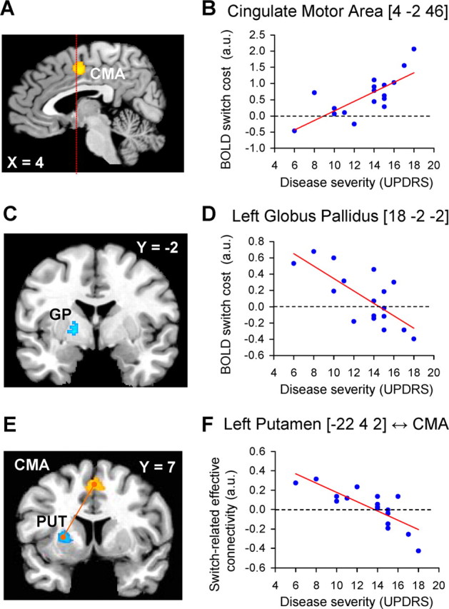
It is well known that the basal ganglia are involved in switching between movement sequences. Here we test the hypothesis that this contribution is an instance of a more general role of the basal ganglia in selecting actions that deviate from the context defined by the recent motor history, even when there is no sequential structure to learn or implement. We investigated the effect of striatal dopamine depletion [in Parkinson's disease (PD)] on the ability to switch between independent action plans. PD patients with markedly lateralized signs performed a hand laterality judgment task that involved action selection of their most and least affected hand. Trials where patients selected the same (repeat) or the alternative (switch) hand as in a previous trial were compared, and this was done separately for the most and least affected hand. Behaviorally, PD patients showed switch-costs that were specific to the most affected hand and that increased with disease severity. Functional magnetic resonance imaging (fMRI) showed that this behavioral effect was related to the state of the frontostriatal system: as disease severity increased, contributions of the basal ganglia to the selection process and their effective connectivity with the medial frontal cortex (MFC) decreased, whereas involvement of the MFC increased. We conclude that the basal ganglia are important for rapidly switching toward novel motor plans even when there is no sequential structure to learn or implement. The enhanced MFC activity may result either from reduced focusing abilities of the basal ganglia or from compensatory processes.


Figures

Figure 1.
Figure 1.
Task setup (experiments 1 and 2). For each trial, subjects had to judge whether a visually presented drawing showed a left or right hand. The top panel illustrates two representative stimuli, one for each hand laterality. Crucially, for each trial we considered whether the previous stimulus had the same or a different laterality than the currently displayed hand drawing. Accordingly, we analyzed the effect of factors hand (2 levels: left or right) and hand-order (2 levels: repeat or switch) on reaction times and cerebral activity. The intertrial interval (ITI) between the offset of one trial (i.e., the response) and the onset of the next (i.e., the presentation of the stimulus) randomly varied between 1.5 and 2.5 s, creating a pseudorandom jitter between successive trials.
Figure 2.
Figure 2.
Behavioral performance (experiment 1). The bars show the time that patients used to solve the hand-laterality judgment task (RT, in milliseconds; mean ± SEM). RTs for repeat (black bars) and switch trials (gray bars) are shown separately according to hand laterality (least affected, most affected). A shows RTs from 17 PD patients during fMRI scanning. B shows how the size of the behavioral switch cost for the most affected hand (on the y-axis, calculated as the difference between the averaged log-transformed RT for switch trials and repeat trials) increases linearly with disease severity (for the most affected side, on the x-axis). Each dot represents one patient. * and NS indicate statistically significant and nonsignificant effects, respectively.
Figure 3.
Figure 3.
Switch- and error-related brain activity. The left column shows the anatomical distribution of switch- and error-related activity, the middle column shows the effects size of switch-related cerebral responses and the right column shows the effects size of error-related responses. A–C, Brain regions in which cerebral activity increased during hand-switch trials (compared with hand-repeat trials), specifically for the most affected hand (compared with the least affected hand). D–F, Brain regions in which cerebral activity increased during error trials (compared with correct trials). G–I, Brain regions that were sensitive to both switch- and error-related effects [conjunction analysis of the overlap between switch-related effects (A–C) and error-related effects (D–F) (Nichols et al., 2005)]. These results show a double dissociation in the response profiles of the CMA and the ACC to switching and error processing: the CMA [the posterior part of the switch-related cluster within the MFC (A)] responded only during switch trials (B), but not during error trials (C). In contrast, the ACC [the anterior part of the error-related cluster within the MFC (D)] responded only during error trials (F), but not switch trials (E). The pre-SMA (G) showed both switch- (H) and error-related activity (I). The statistical parametric maps (A, D, G) represent the results of a random effects analysis, shown at an uncorrected threshold of p < 0.001 (for graphical purposes), and superimposed on sagittal sections of a representative brain of the Montreal Neurological Institute series. The histograms (B, E, H and C, F, I) show the mean (± SEM) parameter estimates from this random effects analysis.
Figure 4.
Figure 4.
Correlation between cerebral effects and disease severity. A, C, E show the anatomical distribution of switch-related responses that were modulated by clinical disease severity. B, D, F show the relationship between switch-related activity and disease severity. Switch-related responses and disease severity are shown for the most affected side. A, B, Brain regions in which switch-related activity increased as a function of disease severity. C, D, Brain regions in which switch-related activity decreased as a function of disease severity. E, F show that switch-related interregional coupling between the CMA (E, in orange) and the left putamen/GP (E, in cyan) decreased as a function of disease severity. In B and D, each dot represents switch-related activity for one patient. In E, each dot represents the change in correlation between responses in the left putamen and the CMA during hand-switch trials (compared with hand-repeat trials) for one patient. The statistical parametric maps (A, C, E) represent the results of a random effects analysis, shown at an uncorrected threshold of p < 0.01 (for graphical purposes), and superimposed on sagittal and coronal sections of a representative brain of the Montreal Neurological Institute series. The left side of the figure shows the left side of the brain.

References

    1. Akkal D, Dum RP, Strick PL. Supplementary motor area and presupplementary motor area: targets of basal ganglia and cerebellar output. J Neurosci. 2007;27:10659–10673. - PMC - PubMed
    1. Behrens TE, Woolrich MW, Walton ME, Rushworth MF. Learning the value of information in an uncertain world. Nat Neurosci. 2007;10:1214–1221. - PubMed
    1. Benecke R, Rothwell JC, Dick JP, Day BL, Marsden CD. Disturbance of sequential movements in patients with Parkinson's disease. Brain. 1987;110:361–379. - PubMed
    1. Brown RG, Marsden CD. Internal versus external cues and the control of attention in Parkinson's disease. Brain. 1988;111:323–345. - PubMed
    1. Carbon M, Felice Ghilardi M, Dhawan V, Eidelberg D. Correlates of movement initiation and velocity in Parkinson's disease: A longitudinal PET study. Neuroimage. 2007;34:361–370. - PMC - PubMed

Publication types