Case Reports
. 2009 May 14;360(20):2099-107.
doi: 10.1056/NEJMoa0806326.

Fatal case of deer tick virus encephalitis

Affiliations
Case Reports

Fatal case of deer tick virus encephalitis

Norma P Tavakoli et al. N Engl J Med. .

Abstract

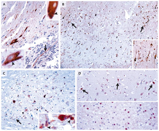
Deer tick virus is related to Powassan virus, a tickborne encephalitis virus. A 62-year-old man presented with a meningoencephalitis syndrome and eventually died. Analyses of tissue samples obtained during surgery and at autopsy revealed a widespread necrotizing meningoencephalitis. Nucleic acid was extracted from formalin-fixed tissue, and the presence of deer tick virus was verified on a flavivirus-specific polymerase-chain-reaction (PCR) assay, followed by sequence confirmation. Immunohistochemical analysis with antisera specific for deer tick virus identified numerous immunoreactive neurons, with prominent involvement of large neurons in the brain stem, cerebellum, basal ganglia, thalamus, and spinal cord. This case demonstrates that deer tick virus can be a cause of fatal encephalitis.


Conflict of interest statement

No potential conflict of interest relevant to this article was reported.

Figures

Figure 1
Figure 1. Magnetic Resonance Imaging (MRI) of the Brain on Hospital Admission
MRI scanning that was performed on hospital day 1 revealed abnormal T2-weighted signaling in the superior cerebellum (Panel A, arrow) and abnormal T2-weighted fluid-attenuated inversion recovery images with hyperintensities in the cerebellum and left pons (Panel B, arrows) and in the bilateral basal ganglia (Panel C). The superior cerebellum was bright on diffusion-weighted imaging (Panel D) and dark on apparent-diffusion-coefficient sequences, which suggested an ischemic process.
Figure 2
Figure 2. Histologic Findings at Autopsy
In Panel A, microglial nodules and lymphocytic infiltrates in the pons are visible in basal pontine nuclei (arrowheads), with less prominent involvement of descending fiber tracts (arrow) and pontocerebellar fibers. In Panel B, confluent foci of parenchymal necrosis can be seen in pontine basal nuclei. In Panel C, CD8+ immunostaining of the basis pontis shows a cytotoxic T-cell infiltrate and a close association with surviving neurons (arrows). In Panel D, nearly complete neuronal loss is seen in the substantia nigra with rare surviving neurons (arrows); in the inset, an eosinophilic dying neuron and remaining neuromelanin pigment are engulfed in macrophages or free in the parenchyma (arrowheads). In Panel E, phosphoglucomutase 1 immunostaining of lumbar spinal cord shows marked infiltration by microglia–macrophages and in the anterior horn and focal microglial nodules in the lateral corticospinal tract (arrow) and posterior column (arrowhead). In Panels A, B, and D, paraffin sections were stained with hematoxylin and eosin.
Figure 3
Figure 3. Phylogenetic Tree Showing the Relationship between the Virus (DT-NY-07) Detected in Tissue Sections from the Brain of the Patient and Other Powassan Viruses
This phylogenetic tree was constructed from 2304 nucleotide sequences of the NS5 region. GenBank accession numbers are in parentheses. The evolutionary history was inferred with the use of the neighbor-joining method. The optimal tree with the sum of branch length equaling 0.60849794 is shown. The percentage of replicate trees in which the associated taxa are clustered together in the bootstrap test (1000 replicates) is shown next to each branch. The tree is drawn to scale, with branch lengths in the same units as those of the evolutionary distances used to construct the phylogenetic tree. To root the dendrogram, louping ill virus was used as the outgroup. The evolutionary distances were computed with the use of the maximum-composite-likelihood method and are expressed in the units of the number of base substitutions per site. All positions containing gaps and missing data were eliminated from the data set. Phylogenetic analyses were conducted with the use of Molecular Evolutionary Genetics Analysis (MEGA) software, version 4.0.
Figure 4
Figure 4. Immunohistochemical Analysis with Deer Tick Virus Antiserum Samples
Paraffin sections of cerebellar samples obtained from the patient on biopsy (Panel A) and samples from the hippocampus (Panel B), pons (Panel C), and temporal cortex (Panel D) obtained at autopsy were stained either with antibody against whole deer tick virus (Panel A, upper inset; and Panels B and C) or with antibody against recombinant deer tick virus E protein (rEDTV) (Panel A; Panel A, lower inset; and Panel D). In Panel A, in the cerebellar-biopsy sample, both types of antiserum recognized surviving Purkinje cells, with prominent filling of their dendrites in the molecular layer and occasional identification of axons in the granule-cell layer (arrow); in the insets, several Purkinje cells were identified with immunoreactive granular-to-tubular profiles (arrowheads). In Panel B, many hippocampal pyramidal neurons were immunolabeled in a segmental distribution (in area surrounding arrows), with prominent decoration of apical and basal processes (inset). In Panel C, many surviving immunolabeled neurons in the basis pontis are visible. The whole deer tick virus antibody also recognized viral antigens engulfed in macrophages (arrow; inset, arrowheads), whereas the rEDTV antibody did not have such recognition. In Panel D, in temporal cortex, immunoreactive neurons that were not associated with inflammatory reaction were occasionally identified (upper panel, arrows). In the temporal white matter, a focus of labeled cells consistent with oligodendrocytes was seen (lower panel). (For more details, see Fig. 5 and 6 in the Supplementary Appendix.)

References

    1. Telford SR, III, Armstrong PM, Katavolos P, et al. A new tick-borne encephalitis-like virus infecting New England deer ticks, Ixodes dammini. Emerg Infect Dis. 1997;3:165–70. - PMC - PubMed
    1. Kuno G, Artsob H, Karabatos N, Tsuchiya KR, Chang GJJ. Genomic sequencing of deer tick virus and phylogeny of Powassan-related viruses of North America. Am J Trop Med Hyg. 2001;65:671–6. - PubMed
    1. Beasley DWC, Suderman MT, Holbrook MR, Barrett ADT. Nucleotide sequencing and serological evidence that the recently recognized deer tick virus is a genotype of Powassan virus. Virus Res. 2001;79:81–9. - PubMed
    1. Ebel GD, Spielman A, Telford SR., III Phylogeny of North American Powassan virus. J Gen Virol. 2001;82:1657–65. - PubMed
    1. Charrel RN, Attoui H, Butenko AM, et al. Tick-borne virus diseases of human interest in Europe. Clin Microbiol Infect. 2004;10:1040–55. - PubMed

Publication types

Substances