p53 regulates mtDNA copy number and mitocheckpoint pathway
- PMID: 19439913
- PMCID: PMC2687143
- DOI: 10.4103/1477-3163.50893
p53 regulates mtDNA copy number and mitocheckpoint pathway
Abstract
Background: We previously hypothesized a role for mitochondria damage checkpoint (mito-checkpoint) in maintaining the mitochondrial integrity of cells. Consistent with this hypothesis, defects in mitochondria have been demonstrated to cause genetic and epigenetic changes in the nuclear DNA, resistance to cell-death and tumorigenesis. In this paper, we describe that defects in mitochondria arising from the inhibition of mitochondrial oxidative phosphorylation (mtOXPHOS) induce cell cycle arrest, a response similar to the DNA damage checkpoint response.
Materials and methods: Primary mouse embryonic fibroblasts obtained from p53 wild-type and p53-deficient mouse embryos (p53 -/-) were treated with inhibitors of electron transport chain and cell cycle analysis, ROS production, mitochondrial content analysis and immunoblotting was performed. The expression of p53R2 was also measured by real time quantitative PCR.
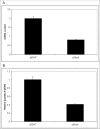
Results: We determined that, while p53 +/+ cells arrest in the cell cycle, p53 -/- cells continued to divide after exposure to mitochondrial inhibitors, showing that p53 plays an important role in the S-phase delay in the cell cycle. p53 is translocated to mitochondria after mtOXPHOS inhibition. Our study also revealed that p53-dependent induction of reactive oxygen species acts as a major signal triggering a mito-checkpoint response. Furthermore our study revealed that loss of p53 results in down regulation of p53R2 that contributes to depletion of mtDNA in primary MEF cells.
Conclusions: Our study suggests that p53 1) functions as mito-checkpoint protein and 2) regulates mtDNA copy number and mitochondrial biogenesis. We describe a conceptual organization of the mito-checkpoint pathway in which identified roles of p53 in mitochondria are incorporated.
Figures


References
-
- Burke DJ. Complexity in the spindle checkpoint. Curr Opin Genet Dev. 2000;10:26–31. - PubMed
-
- Paulovich AG, Toczyski DP, Hartwell LH. When checkpoints fail. Cell. 1997;88:315–21. - PubMed
-
- Stewart ZA, Pietenpol JA. p53 Signaling and cell cycle checkpoints. Chem Res Toxicol. 2001;14:243–2,63. - PubMed
-
- Murray AW. The genetics of cell cycle checkpoints. Curr Opin Genet Dev. 1995;5:5–11. - PubMed
-
- Hartwell LH, Kastan MB. Cell cycle control and cancer. Science. 1994;266:1821–8. - PubMed
Grants and funding
LinkOut - more resources
Full Text Sources
Molecular Biology Databases
Research Materials
Miscellaneous

