

Review
doi: 10.1038/nrc2657.
p21 in cancer: intricate networks and multiple activities
Affiliations
- PMID: 19440234
- PMCID: PMC2722839
- DOI: 10.1038/nrc2657
Item in Clipboard
Review
p21 in cancer: intricate networks and multiple activities
Nat Rev Cancer.
2009 Jun.
Abstract
One of the main engines that drives cellular transformation is the loss of proper control of the mammalian cell cycle. The cyclin-dependent kinase inhibitor p21 (also known as p21WAF1/Cip1) promotes cell cycle arrest in response to many stimuli. It is well positioned to function as both a sensor and an effector of multiple anti-proliferative signals. This Review focuses on recent advances in our understanding of the regulation of p21 and its biological functions with emphasis on its p53-independent tumour suppressor activities and paradoxical tumour-promoting activities, and their implications in cancer.
Figures
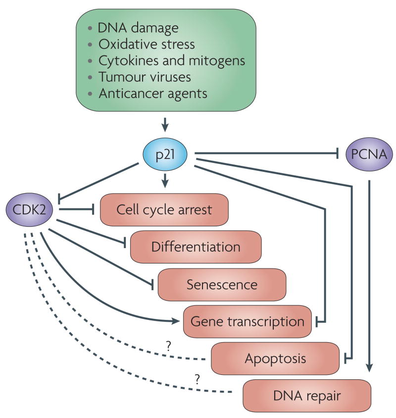
p21 responds to a variety of stimuli to promote growth-inhibitory activities that depend primarily on its ability to inhibit the kinase activity of cyclin-dependent kinase 2 (CDK2). p21-induced cell cycle arrest also depends on its ability to inhibit CDK1. p21 can inhibit cellular proliferation independent of CDK2 inhibition by inhibiting proliferating cell nuclear antigen (PCNA), which is required for S phase progression. Some of the anti-proliferative activities of p21 rely on its multiple protein–protein interactions and its ability to regulate gene transcription. The various physiological responses triggered by p21 are interconnected. For example, cell cycle arrest induced by p21 promotes DNA repair by allowing sufficient time for the damaged DNA to be repaired before it is passed to daughter cells and is a major route by which p21 exerts its anti-apoptotic activities. Similarly, the ability of p21 to regulate gene expression is important in promoting cellular senescence. The effect of p21 on gene transcription is generally inhibitory, but p21 can also activate gene transcription under certain conditions. The role of p21 in promoting DNA damage-induced and p53-dependent cell cycle arrest is well established and not the focus of this Review, and its role in mediating cellular responses to oxidative stress is well described.
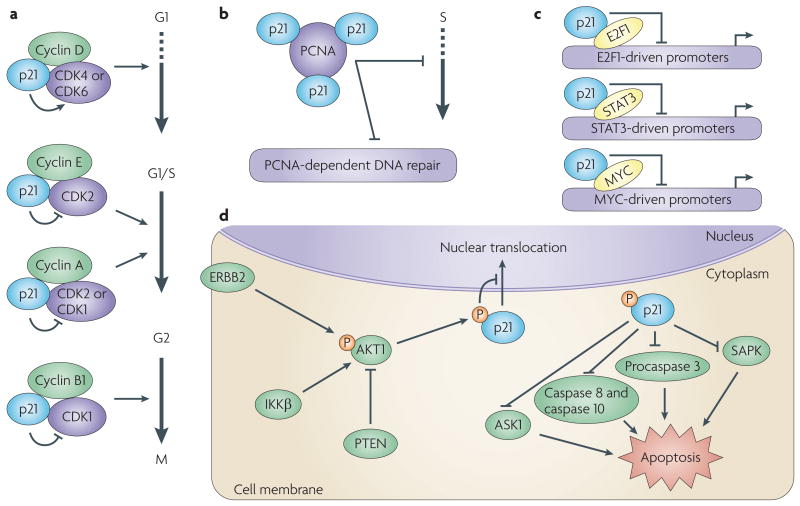
The figure shows activities of p21 in the nucleus and cytoplasm. a | Under certain conditions, p21 promotes the kinase activity of cyclin-dependent kinase 4 (CDK4) or CDK6 in complex with cyclin D, thus promoting progression through G1 (REF. 163). p21 inhibits CDK2–cyclin E, with the consequent inhibition of CDK2-dependent phosphorylation of RB and the sequestration of E2F1, thus inhibiting E2F1-dependent gene transcription and progression into and through S phase. p21 also inhibits the kinase activity of CDK2–cyclin A and CDK1–cyclin A, which are required for progression through S phase and into G2 respectively. Additionally, p21 inhibits the kinase activity of CDK1–cyclin B1, thus inhibiting progression through G2 and G2/M. b | Through its carboxyl-terminal domain, p21 binds to and inhibits proliferative cell nuclear antigen (PCNA), thereby inhibiting processive DNA synthesis and modulating PCNA-dependent DNA repair pathways. c | p21 can inhibit the transcriptional activity of the transcription factors E2F1, STAT3 (signal transducer and activator of transcription 3) and MYC through direct binding and inhibition of their transactivation activity. This accounts for some of the anti-apoptotic effects of p21, which may contribute to its oncogenic activity. d | p21 phosphorylation at Thr145 by activated AKT1 (also known as PKB) downstream of ERBB2 (a member of the epidermal growth factor receptor family of receptor tyrosine kinases) or IKKβ (inhibitor of nuclear factor-κB kinase-β) signalling prevents the nuclear translocation of p21 (REFS 48,129,131). Cytoplasmic p21 exhibits anti-apoptotic activity owing to the inhibition of proteins involved in apoptosis. Whether the phosphorylation of p21 by AKT1 only functions to retain p21 in the cytoplasm or is also required for its cytoplasmic activities is not clear. ASK1, apoptosis signal-regulating kinase 1, also known as MAP3K5; SAPK, stress-activated protein kinase.
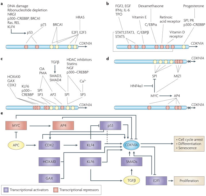
Multiple signals and factors regulate transcription from the CDKN1A promoter. The four SP1-binding sites (yellow circles) in the proximal region of the CDKN1A promoter provide a relative reference for the position of other cis-elements (orange circles). a | Transcriptional activation of CDKN1A in response to a variety of stimuli, including DNA damage, are dependent on p53 and its family member p73. HRAS- and BRCA1-induced CDKN1A transcription, mediated by p53-dependent and p53-independent mechanisms, are also shown. b | Transcriptional activation of CDKN1A by growth factor and nuclear receptors. c | Activation of CDKN1A transcription by transcription factors and chemicals including anticancer agents (such as the histone acetyltransferase (HDAC) inhibitors) and drugs with anti-proliferative activity (such as statins). d | MYC represses CDKN1A transcription by binding to and inhibiting SP1 (REF. 189), and this can be alleviated by the binding of the ligand-independent nuclear receptor hepatocyte nuclear factor 4α1 (HNF4α1) to SP1 (REF. 190). In response to DNA damage, MYC is recruited to the CDKN1A promoter by MIZ1, and forms a ternary complex with the DNA methyltransferase DNMT3a, which represses CDKN1A transcription. Additionally, AP4, a basic helix–loop–helix protein and a transcriptional target of MYC, represses the CDKN1A promoter through binding to four proximal E-box motifs independently of MIZ1, SP1 or SP3 (REF. 107). e | The CDKN1A transcriptional circuitry is shown, comprising transcription factors that upregulate (purple boxes) or downregulate (orange boxes) CDKN1A transcription under various conditions leading to growth arrest, differentiation or cellular senescence. Several of these factors function in transcriptional networks. APC, adenomatous polyposis coli; C/EBPα, CCAAT/enhancer binding protein-α; CREBBP, CREB binding protein; FGF2, fibroblast growth factor 2; GAX, also known as MOX2; HOXA10, homeobox A10; IFNγ, interferon-γ; IL-6, interleukin 6; KLF4, Krüppel-like factor 4; NGF, nerve growth factor; NRG1, neuregulin; OA, okadaic acid; PMA, phorbol-12-myristate-13-acetate; PR, progesterone receptor; STAT, signal transducer and activator of transcription; TGFβ, transforming growth factor-β; TPO, thrombopoietin.
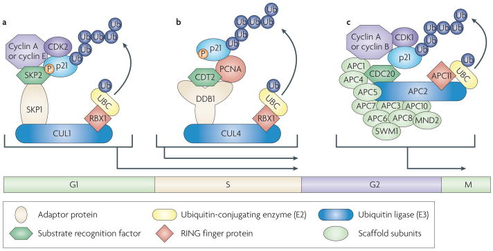
The figure shows ubiquitin-mediated proteolysis of p21 during an unperturbed cell cycle. a | The SCFSKP2 (SKP1–CUL1–SKP2) E3 ubiquitin ligase complex promotes the ubiquitylation and degradation of p21 that is phosphorylated by cyclin-dependent kinase 2 (CDK2) at Ser130 at the G1/S transition and during S phase of the cell cycle, thus selectively de-repressing CDK2 kinase,–. b | A second CRL (cullin-RING ligase), the CRL4CDT2 (CUL4A or CUL4B–DDB1–CDT2 (DDB1 is DNA damage-binding protein 1)) E3 ubiqutin ligase complex, targets p21 for ubiquitin-dependent proteolysis specifically in S phase only when it is bound to proliferating cell nuclear antigen (PCNA),,. The CRL4CDT2 ubiquitin ligase also targets p21 for degradation in response to low and moderate doses of ultraviolet irradiation, and after ionizing radiation in a PCNA-dependent fashion. c | The degradation of p21 during G2/M is carried out by the APC/CCDC20 (anaphase-promoting complex (APC)–cell division cycle 20) E3 ubiquitin ligase complex, which recognizes CDK1–cyclin A- and CDK1–cyclin B-bound forms of p21, and is important for CDK1 activity necessary for mitosis. The inhibition of APC/CCDC20 during spindle checkpoint activation results in the stabilization of p21, which inhibits CDK1 kinase activity and prevents premature entry of cells into mitosis. SKP2, CDT2 and CDC20 function as substrate recognition factors for the respective ubiquitin ligase complexes and bridge p21 to the rest of the E3 ligase. RBX1, RING box protein 1; UBC, ubiquitin-conjugating enzyme.
References
-
- Bartek J, Lukas J. DNA damage checkpoints: from initiation to recovery or adaptation. Curr Opin Cell Biol. 2007;19:238–245. - PubMed
-
- Eastman A. Cell cycle checkpoints and their impact on anticancer therapeutic strategies. J Cell Biochem. 2004;91:223–231. - PubMed
-
- Deng C, Zhang P, Harper JW, Elledge SJ, Leder P. Mice lacking p21CIP1/WAF1 undergo normal development, but are defective in G1 checkpoint control. Cell. 1995;82:675–684. - PubMed
-
- Brugarolas J, et al. Radiation-induced cell cycle arrest compromised by p21 deficiency. Nature. 1995;377:552–557. - PubMed
Publication types
MeSH terms
Substances
Grants and funding
LinkOut - more resources
Full Text Sources
Other Literature Sources
Research Materials
Miscellaneous
