. 2009;4(5):e5547.
doi: 10.1371/journal.pone.0005547. Epub 2009 May 14.

Mucosal immunization of cynomolgus macaques with the VSVDeltaG/ZEBOVGP vaccine stimulates strong ebola GP-specific immune responses

Affiliations

Mucosal immunization of cynomolgus macaques with the VSVDeltaG/ZEBOVGP vaccine stimulates strong ebola GP-specific immune responses

Xiangguo Qiu et al. PLoS One. 2009.

Abstract

Background: Zaire ebolavirus (ZEBOV) produces a lethal viral hemorrhagic fever in humans and non-human primates.

Methodology/principal findings: We demonstrate that the VSVDeltaG/ZEBOVGP vaccine given 28 days pre-challenge either intranasally (IN), orally (OR), or intramuscularly (IM) protects non-human primates against a lethal systemic challenge of ZEBOV, and induces cellular and humoral immune responses. We demonstrated that ZEBOVGP-specific T-cell and humoral responses induced in the IN and OR groups, following an immunization and challenge, produced the most IFN-gamma and IL-2 secreting cells, and long term memory responses.

Conclusions/significance: We have shown conclusively that mucosal immunization can protect from systemic ZEBOV challenge and that mucosal delivery, particularly IN immunization, seems to be more potent than IM injection in the immune parameters we have tested. Mucosal immunization would be a huge benefit in any emergency mass vaccination campaign during a natural outbreak, or following intentional release, or for mucosal immunization of great apes in the wild.


Conflict of interest statement

Competing Interests: The authors have declared that no competing interests exist.

Figures

Figure 1
Figure 1. Survival of vaccinated cynomolgus macaques.
A) Flow chart of experimental design. Arrows indicate vaccination and challenge date. Tick marks indicate sampling days. B) Kaplan-Meier survival curve for cynomolgus macaques immunized by different routes and challenged with Zaire ebolavirus. Cynomolgus macaques were immunized orally (OR; n = 4), intranasally (IN; n = 4) or intramuscularly (IM; n = 2) with 2×10⁷ PFU of VSVΔG/ZEBOVGP, or injected intramuscularly with VSVΔG/MARVGP (control; n = 2). All animals were challenged 28 days later with 1000 PFU ZEBOV. The animals were scored daily for fever, macular rashes, lethargy, and unresponsiveness.
Figure 2
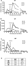
Figure 2. Strong humoral IgM, IgA, IgG responses are induced in the IM, OR, and IN immunization routes.
A ZEBOV VLP ELISA was utilized to determine the ZEBOV-specific IgA (a), IgG (b), IgM (c) titres in post-vaccination and post-challenge sera from cynomolgus macaques immunized either orally (OR; n = 4), intranasally (IN; n = 4) or intramuscularly (IM; n = 2). Titres are presented as endpoint dilutions of the average value per group. Table (d) illustrates fold increases in peak titres post-challenge compared to post-vaccination for IgA and IgG. IgM is not shown as there were no increases post-challenge.
Figure 3
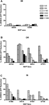
Figure 3. Neutralizing antibodies generated by the various immunization routes.
ZEBOV GP-specific neutralizing antibodies in the sera from NHPs vaccinated either (A) intramuscularly (IM), (B) orally (OR), or (C) intranasally (IN), were investigated for their ability to inhibit infection by ZEBOV-GFP. NHP sera from days 0 and 21 post-vaccination were incubated for 1 hour with ZEBOV-GFP before being added to a monolayer of Vero cells. The positive control was ZEBOV-GFP in DMEM without sera. The level of GFP fluorescence of ZEBOV-GFP infected cells was determined by flow cytometry. The percent reduction of infection by ZEBOV-GFP was calculated as follows: % ZEBOV-GFP reduction = (1−(Test samples/Positive control))×100). The day 21 results for each NHP are displayed and have been normalized by subtracting each NHP's day 0 results from the day 21 results. The numbers in the legend represent the dilution of the NHP sera that was added to ZEBOV-GFP.
Figure 4
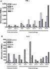
Figure 4. PBMCs from immunized animals produce IFN-γ or IL-2 in response to ZEBOVGP peptides.
ZEBOVGP-specific IFN-γ or IL-2 secreting cells from PBMCs were detected by either an (A) IFN-γ or (B) IL-2 ELISPOT assay. PBMCs were obtained from cynomolgus macaques immunized with VSVΔG/ZEBOVGP either orally (OR; n = 4), intranasally (IN; n = 4), or intramuscularly (IM; n = 2). The control NHPs (n = 2) were immunized IM with the heterologous VSVΔG/MARVGP. PBMCs were incubated with 1.5 µg/ml of peptides spanning the entire ZEBOV glycoprotein. Bars represent the average number of ZEBOVGP-specific IFN-γ or IL-2 secreting cells detected in each immunized group.
Figure 5
Figure 5. Absolute white blood cell numbers do not decrease in immunized animals after Zaire ebolavirus challenge.
Whole blood from cynomolgus macaques immunized either orally (OR; n = 4), intranasally (IN; n = 4) or intramuscularly (IM; n = 2) was stained for lympocytes (CD3+, CD4+, CD8+), monocytes (CD14+). Control animals (n = 2) received non protective VSVΔG/MARVGP. The absolute numbers in blood was determined for the each of the monkeys by flow cytometry on days 0, 3 and 6 post-challenge with ZEBOV. The average for each inoculation route is represented.
Figure 6
Figure 6. ZEBOVGP-specific CD4+ and CD8+ functional memory responses in PBMCs.
Flow cytometry was utilized to evaluate the long term CD4+ (A) and CD8+ (B) ZEBOVGP-specific functional memory responses of freshly isolated PBMCs incubated with media or ZEBOVGP peptides at 6 months post-vaccination. The average for each inoculation route (IM = intramuscular n = 2, OR = orally n = 4, IN = intranasally n = 4) is represented. The CD45RA population denotes the memory lymphocytes, while CD45RA+ represents the naïve and effector lymphocytes. The IFN-γ response was evaluated after 3 days by intracellular cytokine staining. The background from the media sample was subtracted from the peptide stimulated sample, and the results are shown for each monkey with the average for each group being represented with a bar. The ability of the PBMCs to proliferate in response to the GP peptide was determined by staining the PBMCs with CFSE and then looking for loss of CFSE 6 days later as a measure of proliferation. The percent of CFSE cells for each memory population is shown for each monkey after subtracting the media sample, and the average for each group is represented by a bar.

References

    1. Feldmann H, Jones S, Klenk HD, Schnittler HJ. Ebola virus: From discovery to vaccine. Nat Rev Immunol. 2003;3(8):677–685. - PubMed
    1. Geisbert TW, Jahrling PB. Exotic emerging viral diseases: Progress and challenges. Nat Med. 2004;10(12 Suppl):S110–21. - PubMed
    1. Mason C. The strains of ebola. CMAJ. 2008;178(10):1266–1267. - PMC - PubMed
    1. [Anonymous] Outbreak news. ebola haemorrhagic fever, uganda–end of the outbreak. Wkly Epidemiol Rec. 2008;83(10):89–90. - PubMed
    1. Towner JS, Sealy TK, Khristova ML, Albarino CG, Conlan S, et al. Newly discovered ebola virus associated with hemorrhagic fever outbreak in uganda. PLoS Pathog. 2008;4(11):e1000212. - PMC - PubMed

Publication types

MeSH terms