. 2009 Apr;117(4):515-21.
doi: 10.1289/ehp.11870. Epub 2008 Dec 1.

Time- and dose-related effects of di-(2-ethylhexyl) phthalate and its main metabolites on the function of the rat fetal testis in vitro

Affiliations

Time- and dose-related effects of di-(2-ethylhexyl) phthalate and its main metabolites on the function of the rat fetal testis in vitro

François Chauvigné et al. Environ Health Perspect. 2009 Apr.

Abstract

Background: Endocrine-disrupting effects of phthalates are understood primarily from in utero exposures within the fetal rat testis. Nevertheless, their path of action, dose-response character, and cellular target(s) within the fetal testis are not known.

Objectives: In this study we investigated the effects of di-(2-ethylhexyl) phthalate (DEHP), mono-(2-ethylhexyl) phthalate (MEHP), and several of their metabolites on the development of organo-cultured testes from rat fetus.

Methods: We removed testes from 14.5-day-old rat fetuses and cultured them for 1-3 days with or without DEHP, MEHP, and the metabolites.

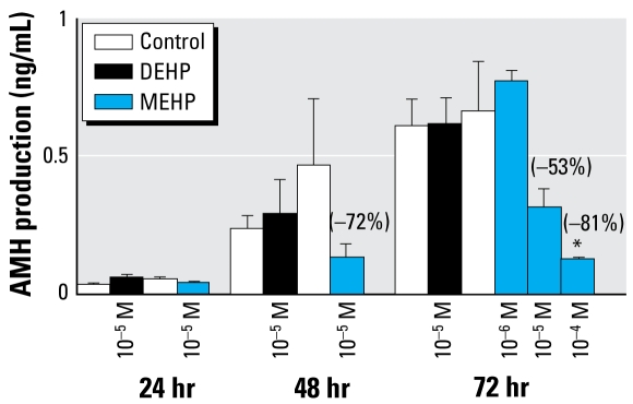
Results: DEHP (10(-5) M) produced a proandrogenic effect after 3 days of culture, whereas MEHP disrupted testis morphology and function. Leydig cells were the first affected by MEHP, with a number of them being inappropriately located within some seminiferous tubules. Additionally, we found a time- and dose-dependent reduction of testosterone. By 48 hr, gonocyte proliferation had decreased, whereas apoptosis increased. Sertoli cell number was unaffected, although some cells appeared vacuolated, and production of anti-Müllerian hormone decreased in a time- and dose-dependent manner. The derived metabolite mono-(2-ethyl-5-hydroxyhexyl) phthalate was the only one to cause deleterious effects to the rat fetal testis in vitro.

Conclusion: We hope that this in vitro method will facilitate the study of different phthalate esters and other endocrine disruptors for direct testicular effects.

Keywords: androgens; anti-Müllerian hormone; endocrine disruption; explant culture; fetal testis; gonocytes; phthalates.


Figures

Figure 1
Figure 1
Effect of DEHP and MEHP on testicular histology (AD) and the total number of gonocytes (E) after 72 hr of culture beginning on GD14.5 (see “Materials and Methods” for experimental details). (A) Histology of nonexposed (control) fetal testis. (B, C, D) Effect of 10−6 M (B), 10−5 M (C), or 10−4 M (D) MEHP on the histology of seminiferous tubules in fetal rat testes after 72 hr in culture. Gonocytes were immunostained with DDX4/VASA antibody, revealed by AEC (red), and Leydig cells were immunostained with 3β-HSD antibody, revealed by DAB (brown). Scale in (A) also applies to (B–D). Arrows in (B) and (C) indicate holes and degenerative gonocytes within seminiferous tubules. In (D), seminiferous tubules were not recognizable. (E) Effect of DEHP and MEHP on the total number of gonocytes. Responses to DEHP and MEHP were measured by comparing one control testis (DMSO-treated) with the contra lateral testis cultured in medium containing the tested factor. Values are mean ± SE of 3–7 fetuses in at least two independent experiments. The numbers in parentheses indicate the percent decrease relative to the corresponding control. *p < 0.05, and **p < 0.01 by Wilcoxon signed rank tests performed on paired data.
Figure 2
Figure 2
Effect of 10−5 M MEHP on the mitotic index and apoptosis of gonocytes. (A) Quantitative analysis of BrdU incorporation into gonocytes at 48 and 72 hr of culture measured as the percentage of BrdU-positive gonocytes in at least 500 cells (n = 4 fetuses at each time point). (B) Quantitative analysis of apoptotic gonocytes measured as the percentage of TUNEL-positive gonocytes in at least 500 cells (n = 4 fetuses at 48 hr and n = 6 fetuses at 72 hr). The numbers in parentheses indicate the percent decrease (A ) or the factor of increase (B) relative to the corresponding control. *p < 0.05 by Wilcoxon signed rank tests on paired data.
Figure 3
Figure 3
Effect of DEHP and MEHP on Sertoli cell morphology (A–C) and the total number of Sertoli cells (D) in cultured testes. Con, control. (A–C) One testis from each fetus was cultured in control medium (A) and the other in medium supplemented with DEHP (B) or MEHP (C) at 10−5 M. Sertoli cells were immunostained with WT-1 antibody, revealed by DAB (brown). In the MEHP-treated testis (C), some Sertoli cells appeared vacuolated (arrows) and aggregated (arrowheads). (D ) Sertoli cells were counted after 3 days of culture using systematic random sampling accomplished by the CAST-grid stereotaxic system. Values are mean ± SE of 7–8 fetuses from at least two independent experiments analogous to A–C. No significant difference was found in the Sertoli cell number by Wilcoxon signed rank tests of paired data.
Figure 4
Figure 4
Effects of DEHP (10−5 M) and MEHP (10−6 M, 10−5 M, or 10−4 M) on Sertoli cell function revealed by in vitro AMH secretion by GD14.5 fetal rat testes cultured for 24, 48, or 72 hr and measured by ELISA (see “Materials and Methods” for experimental details). Values are mean ± SE of 3–4 fetuses; values in parentheses indicate the decrease relative to control. In the dose–response study at 72 hr, four groups of testes (from different dams and formed randomly) were exposed to 10−5 M DEHP or 10−6 to 10−4 M MEHP, and compared with corresponding control values. A non-parametric ANOVA on paired data with repeated measures indicated that in controls, a statistically significant increase of AMH production occurred over time, and a statistically significant decrease of AMH production occurred with 10−5 M MEHP exposure (p< 0.05). *p < 0.05 by Wilcoxon signed rank tests of paired data.
Figure 5
Figure 5
Effect of DEHP and MEHP on the location (A–C) and number (D) of Leydig cells after 72 hr of culture beginning on GD14.5 (see “Materials and Methods” for experimental details). Con, Control. (A–C) Leydig cells were immuno stained with 3β-HSD antibody and revealed by AEC (red) after treatment with control (A), 10−5 M DEHP (B) or 10−5 M MEHP (C). After MEHP exposure, we observed a low but consistent number of Leydig cells abnormally located within the seminiferous tubules (C, arrows). The scale in (A) also applies to (B–C). (D ) Leydig cells were counted using systematic random sampling accomplished by the CAST-grid stereotaxic system. Values are mean ± SE of 4 fetuses from at least two independent experiments. No significant difference in number was found using Wilcoxon signed rank tests.
Figure 6
Figure 6
Time course (A) and dose response (B) showing effects of DEHP and MEHP on basal testosterone secretion by fetal rat testes cultured beginning on GD14.5. Testosterone was measured by radioimmuno assay. (A) For the time course, one testis from each fetus was cultured in the control medium, and the contralateral testis was cultured in medium containing DEHP or MEHP. One-half of medium was changed every 24 hr. (B) Effects of increasing concentrations of DEHP and MEHP on basal testosterone production after 72 hr of culture. In this case, the control and phthalate-exposed testes did not originate from the same fetuses; the testes of different fetuses and from different dams were assigned at random. In (A), values are mean ± SE of 18–34 fetuses from at least three independent experiments; in (B), values are mean ± SE of 7–14 testes from at least two independent experiments. The values in parentheses indicate the percentage of decrease or increase relative to the corresponding control. *p < 0.05; and **p < 0.01 by Wilcoxon signed rank tests on paired data in (A) and nonparametric ANOVA on unpaired data in ( B).
Figure 7
Figure 7
Time course (A) and dose response (B) showing effects of MEHP and DEHP on basal and LH-stimulated testosterone secretion by fetal rat testes cultured beginning on GD14.5. (A) For the time course, one testis from each fetus was cultured for 27, 51, or 75 hr in control medium or in medium containing MEHP, with media changed every 24 hr. For the last 3 hr of the culture period, medium was supplemented with (+LH) or without (−LH) 100 ng/mL LH (24–27 hr, 48–51 hr, and 72–75 hr). (B) Effects of 10−5 M DEHP and increasing concentrations of MEHP on basal and LH-stimulated testosterone production after 72 hr of culture. In this case, the control and phthalate-exposed testes did not originate from the same fetuses; the testes of different fetuses and from different mothers were assigned at random. In (A), values are mean ± SE of 6–18 fetuses from at least two independent experiments; in (B), values are mean ± SE of 3–10 testes from at least two independent experiments. The values in parentheses indicate the percentage of decrease (−) or increase (+) relative to control. *p < 0.05; and **p < 0.01 by Wilcoxon signed rank tests on paired data in (A) and nonparametric ANOVA on unpaired data in (B). ND, not detectable.
Figure 8
Figure 8
Effects of DEHP metabolites MEHP, 5OH-MEHP, 5OXO-MEHP, or 5CX-MEPP (10−5 M) on the number of gonocytes (A) and on testosterone production (B) in fetal rat testes cultured for 72 hr beginning on GD14.5. One-half of medium was changed every 24 hr for both (A) and (B). (B) Testosterone was measured by radioimmuno assay. In (A), values are mean ± SE of 5–7 fetuses from at least two independent experiments; in (B), values are mean ± SE of 6–20 animals from at least two independent experiments. Values in parentheses indicate the percent decrease relative to corresponding control. *p < 0.05 by Wilcoxon signed rank tests on paired data.

Similar articles

Cited by

References

    1. Abercrombie M. Estimation of nuclear population from microtome sections. Anat Rec. 1946;94:238–248. - PubMed
    1. ATSDR. Toxicological Profile for Diethyl Phthalate. Atlanta, GA: Agency for Toxic Substances and Disease Registry; 2002. [[accessed 18 February 2009]]. Available: http://www.atsdr.cdc.gov/toxprofiles/tp73.html. - PubMed
    1. Barlow NJ, Phillips SL, Wallace DG, Sar M, Gaido KW, Foster PM. Quantitative changes in gene expression in fetal rat testes following exposure to di(n-butyl) phthalate. Toxicol Sci. 2003;73(2):431–441. - PubMed
    1. Becker K, Seiwert M, Angerer J, Heger W, Koch HM, Nagorka R, et al. DEHP metabolites in urine of children and DEHP in house dust. Int J Hyg Environ Health. 2004;207(5):409–417. - PubMed
    1. Boekelheide K, Lee J, Shipp EB, Richburg JH, Li G. Expression of Fas system-related genes in the testis during development and after toxicant exposure. Toxicol Lett. 1998:102–103. 503–508. - PubMed

Publication types

MeSH terms

LinkOut - more resources