. 2009;4(5):e5586.
doi: 10.1371/journal.pone.0005586. Epub 2009 May 18.

Human neural stem cells genetically modified to overexpress Akt1 provide neuroprotection and functional improvement in mouse stroke model

Affiliations

Human neural stem cells genetically modified to overexpress Akt1 provide neuroprotection and functional improvement in mouse stroke model

Hong J Lee et al. PLoS One. 2009.

Abstract

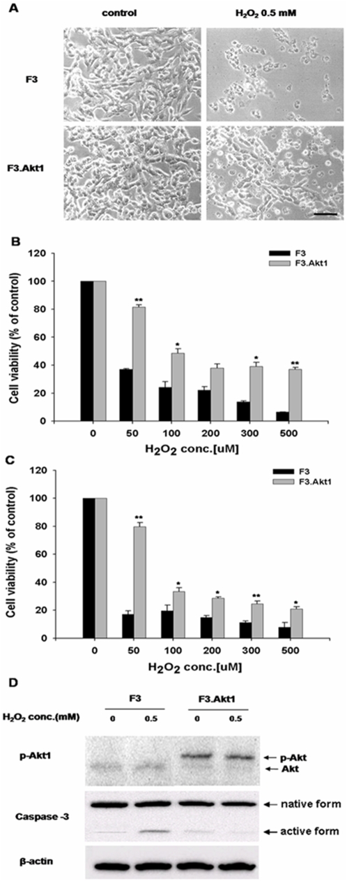
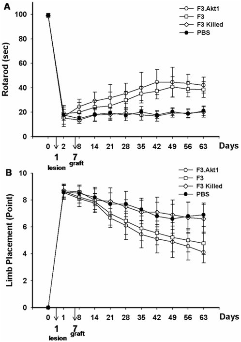
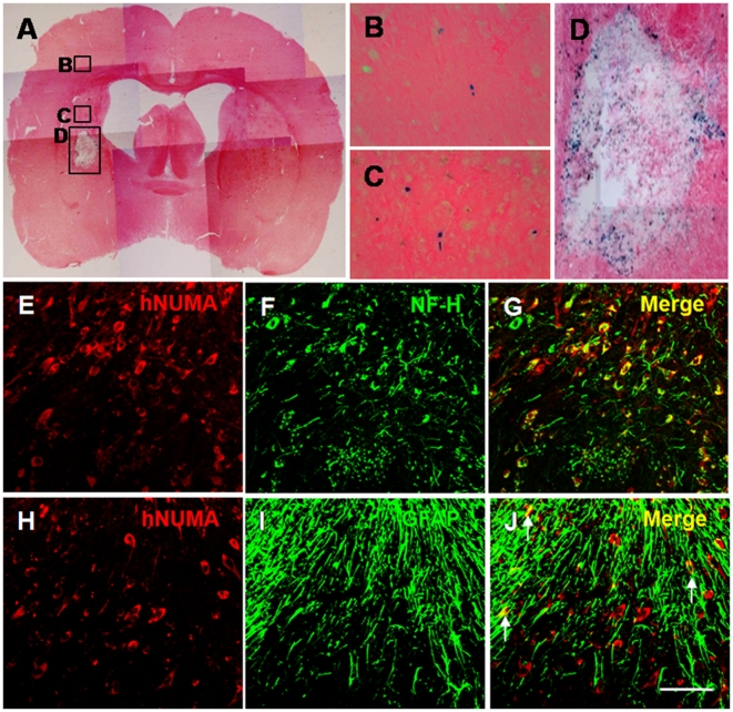
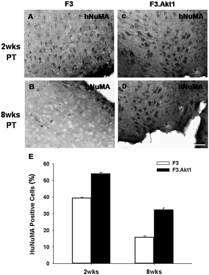
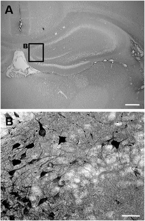
In a previous study, we have shown that human neural stem cells (hNSCs) transplanted in brain of mouse intracerebral hemorrhage (ICH) stroke model selectively migrate to the ICH lesion and induce behavioral recovery. However, low survival rate of grafted hNSCs in the brain precludes long-term therapeutic effect. We hypothesized that hNSCs overexpressing Akt1 transplanted into the lesion site could provide long-term improved survival of hNSCs, and behavioral recovery in mouse ICH model. F3 hNSC was genetically modified with a mouse Akt1 gene using a retroviral vector. F3 hNSCs expressing Akt1 were found to be highly resistant to H(2)O(2)-induced cytotoxicity in vitro. Following transplantation in ICH mouse brain, F3.Akt1 hNSCs induced behavioral improvement and significantly increased cell survival (50-100% increase) at 2 and 8 weeks post-transplantation as compared to parental F3 hNSCs. Brain transplantation of hNSCs overexpressing Akt1 in ICH animals provided functional recovery, and survival and differentiation of grafted hNSCs. These results indicate that the F3.Akt1 human NSCs should be a great value as a cellular source for the cellular therapy in animal models of human neurological disorders including ICH.


Conflict of interest statement

Competing Interests: The authors have declared that no competing interests exist.

Figures

Figure 1
Figure 1. Characterization of human NSC lines.
A: The retroviral vector encoding Akt1 (pLHCX.Akt1) used in the present study for the generation of HB1.F3.Akt1 (F3.Akt1) human neural stem cell (NSC) line. B and C: Phase contrast microscopy of F3 and F3.Akt1 human NSCs. Bar inciates 20 µm. D: Gene expression of cell type-specific markers as studied by RT-PCR in F3 and F3.Akt1 human NSCs. Both of F3 and F3.Akt1 NSC lines express cell type-specific markers Nestin (for neural stem cells), NF-L, NF-M and NF-H (for neurons), GFAP (for astrocytes) and Akt1.
Figure 2
Figure 2. Cell viability increases to H2O2–induced oxydative stress conditions and Akt1 phosphorylation in F3.Akt1 human NSCs.
A: Phase contrast microscopy of F3 and F3.Akt1 human NSCs following exposure to 0.5 mM H2O2 for 6 hr. F3.Akt1 NSCs survived well as compared to the parental F3 NSCs. B and C: F3.Akt1 NSCs were found to show resistance to H2O2-induced cell death as compared to control parental F3 NSCs at 6 hr (B) and 24 hr (C) respectively. D: Western blot analyses of protein levels of phopho-Akt1 and caspase-3 enzymes in F3 and F3.Akt1 NSCs following H2O2 treatment. F3.Akt1 NSCs showed an increased level of Akt1 phosphorylation, while the level in activation form of caspase-3 was reduced under the H2O2 treatment (* p<0.05, ** p<0.001).
Figure 3
Figure 3. Behavioral improvement demonstrated in intracerebral hemorrhage (ICH) mice transplanted with F3 or F3.Akt1 human NSCs.
A: Rotarod test. F3.Akt1-transplanted group showed better performance than PBS controls or F3 cell group, killed F3.Akt1 cell group 8 days onward, and these benefits continued up to 8 weeks post-transplantation (* P<0.05). B: In the modified limb placement test, F3.Akt1-transplanted group showed better performance than PBS, F3 or killed F3.Akt1 group (* P<0.05).
Figure 4
Figure 4. LacZ (beta-galactosidase)-labeled F3.Akt1 human NSCs in intracerebral hemorrhage (ICH) mouse brain at 2 weeks post-transplantation.
A: One week after an ICH lesion (an intrastriatal injection of collagenase), LacZ -labeled F3.Akt1 NSCs were transplanted into the cortex overlying the ICH lesion. Two weeks post-transplantation, LacZ-positive F3.Akt1 NSCs were found to migrate extensively into the hemorrhage core and surrounding brain sites. Bar indicates 50 µm. B–D: Higher magnification of indicated areas. Bar indicates 20 µm. E–G: Double immunofluorescent staining of engrafted F3.Akt1 human NSCs in ICH mouse brain 8 weeks post-transplantation. F3.Akt1 NSCs in the lesion sites are found to differentiate into neurons as shown by double staining of human nuclear matrix antigen (hNuMA) and neurofilament protein (NF-H, a neuron specific marker). H–J: F3.Akt1 human NSCs in the lesion sites are found to differentiate into astrocytes (arrows) as shown by double staining of human nuclear matrix antigen (hNuMA) and glial fibrillary acidic protein (GFAP, an astrocyte specific marker). Bar indicates 20 µm.
Figure 5
Figure 5
At 2 weeks post-transplantation (PT) in the hemorrhage core border areas, difference in number of human nuclear matrix antigen (hNuMA)-positive cells between ICH-F3 control group (A) and ICH-F3.Akt1 group (C) is not apparent, but at 8 weeks PT number of hNuMA-positive cells is much higher in F3.Akt1-ICH group (D) as compared to control F3-ICH group (B). Bar indicates 100 µm. E: Percentage of hNuMA-positive cells found in hemorrhage core border area is higher in ICH-F3.Akt1 group as compared to ICH-F3 group at both 2 and 8 weeks post-transplantation.
Figure 6
Figure 6. Survival of grafted F3.Akt1 human NSCs in hippocampus was demonstrated by immunoperoxidase microscopy of hNuMA at 8 weeks post-transplantation (PT).
A: Lower magnification of hippocampus 8 weeks PT. Bar indicates 1 mm. B: F3.Akt1 human NSCs grafted in cortex overlying striatum were found to migrate extensively to hippocampus 8 weeks PT. Bar indicates 50 µm.

References

    1. Gebel JM, Broderick JP. Intracerebral hemorrhage. Neurol Clin. 2000;18:419–438. - PubMed
    1. NINDS ICH Workshop Participants. Priorities for clinical research in intracerebral hemorrhage: report from a National Institute of Neurological Disorders and Stroke workshop. Stroke. 2005;36:23–41. - PubMed
    1. McKay R. Stem cells in the central nervous system. Science. 1997;276:66–71. - PubMed
    1. Gage FH. Mammalian neural stem cells. Science. 2000;287:1433–1438. - PubMed
    1. Kim SU. Human neural stem cells genetically modified for brain repair in neurological disorders. Neuropathology. 2004;24:159–171. - PubMed

Publication types

MeSH terms