

Brain volume abnormalities in major depressive disorder: a meta-analysis of magnetic resonance imaging studies
- PMID: 19441021
- PMCID: PMC6871089
- DOI: 10.1002/hbm.20801
Brain volume abnormalities in major depressive disorder: a meta-analysis of magnetic resonance imaging studies
Abstract
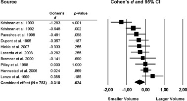
Objective: So far, there have been no attempts to integrate the growing number of all brain volumetric magnetic resonance imaging studies in depression. In this comprehensive meta-analysis the magnitude and extent of brain volume differences between 2,418 patients with major depressive disorder and 1,974 healthy individuals from 64 studies was determined.
Methods: A systematic research was conducted for volumetric magnetic resonance imaging studies of patients with major depressive disorder in relation to healthy control subjects. Studies had to report sufficient data for computation of effect sizes. For each study, the Cohen's d was calculated. All analyses were performed using the random effects model. Additionally, meta-regression analyses were done to explore the effects of potential sources of heterogeneity.
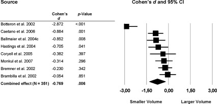
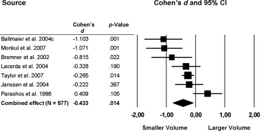
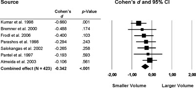
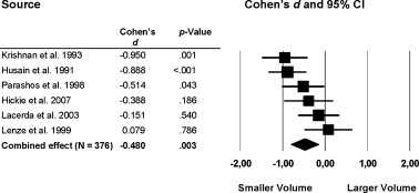
Results: Patients showed large volume reductions in frontal regions, especially in the anterior cingulate and orbitofrontal cortex with smaller reductions in the prefrontal cortex. The hippocampus, the putamen and caudate nucleus showed moderate volume reductions.
Conclusions: This is the first comprehensive meta-analysis in major depressive disorder demonstrating structural brain abnormalities, particularly in those brain areas that are involved in emotion processing and stress-regulation.
Figures

References
-
- Alexander GE, DeLong MR, Strick PL ( 1986): Parallel organization of functionally segregated circuits linking basal ganglia and cortex. Annu Rev Neurosci 9: 357– 381. - PubMed
-
- Alexopoulos GS, Murphy CF, Gunning‐Dixon FM, Latoussakis V, Kanellopoulos D, Klimstra S, Lim KO, Hoptman MJ ( 2008): Microstructural white matter abnormalities and remission of geriatric depression. Am J Psychiatry 165: 238– 244. - PubMed
-
- Almeida OP, Burton EJ, Ferrier N, McKeith IG, O'Brien JT ( 2003): Depression with late onset is associated with right frontal lobe atrophy. Psychol Med 33: 675– 681. - PubMed
-
- Ashburner J, Friston KJ ( 2000): Voxel‐based morphometry—The methods. Neuroimage 11: 805– 821. - PubMed
-
- Ashtari M, Greenwald BS, Kramer‐Ginsberg E, Hu J, Wu H, Patel M, Aupperle P, Pollack S ( 1999): Hippocampal/amygdala volumes in geriatric depression. Psychol Med 29: 629– 638. - PubMed
Publication types
MeSH terms
LinkOut - more resources
Full Text Sources
Other Literature Sources
Medical
