

A geometric analysis of hallux valgus: correlation with clinical assessment of severity
- PMID: 19442286
- PMCID: PMC2694774
- DOI: 10.1186/1757-1146-2-15
A geometric analysis of hallux valgus: correlation with clinical assessment of severity
Abstract
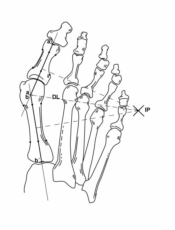
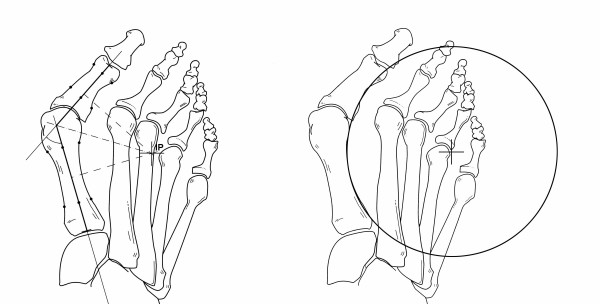
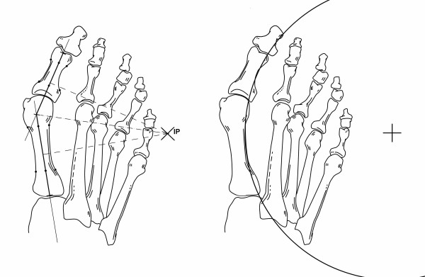
Background: Application of plane geometry to the study of bunion deformity may represent an interesting and novel approach in the research field of hallux valgus. For the purpose of contributing to development of a different perspective in the assessment of hallux valgus, this study was conducted with three objectives: a) to determine the position on the intersection point of the perpendicular bisectors of the longitudinal axes of the first metatarsal and proximal phalanx (IP), b) to correlate the location of this point with hallux valgus deformity according to angular measurements and according to visual assessment of the severity carried out by three independent observers, and c) to assess whether this IP correlated with the radius of the first metatarsophalangeal arc circumference.
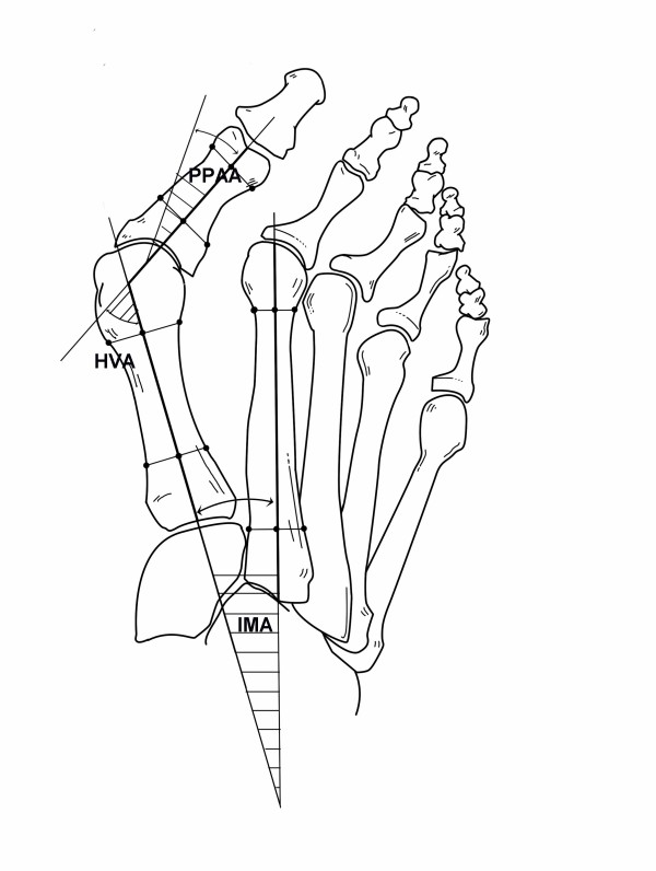
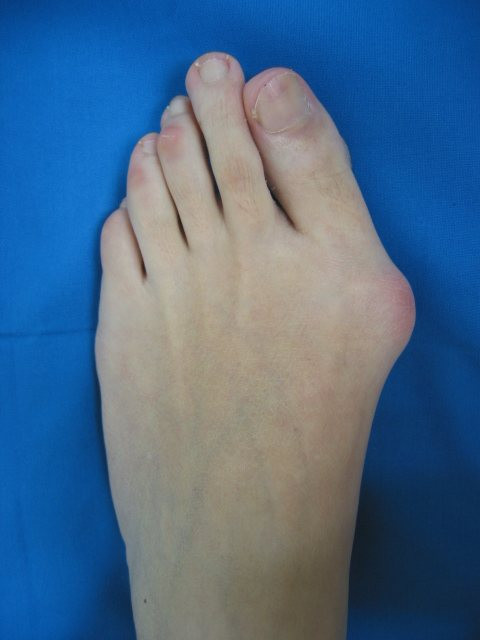
Methods: Measurements evaluated were intermetatarsal angle (IMA), hallux valgus angle (HVA), and proximal phalangeal articular angle (PPAA). The Autocad(R) program computed the location of the IP inside or outside of the foot. Three independent observers rated the severity of hallux valgus in photographs using a 100-mm visual analogue scale (VAS).
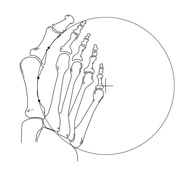
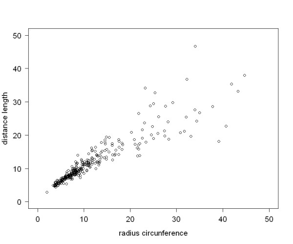
Results: Measurements of all angles except PPAA showed significantly lower values when the IP was located out of the foot more distantly and vice versa, significantly higher values for severe deformities in which the IP was found inside the foot (p < 0.001). The IP correlated significantly with VAS scores and with the length of the radius of the circle that included the first metatarsophalangeal arc circumference (p < 0.001)
Conclusion: The IP is a useful indicator of hallux valgus deformity because correlated significantly with IMA and HVA measurements, VAS scores obtained by visual inspection of the degree of deformity, and location of the center of the first metatarsophalangeal arc circumference.
Figures
References
-
- Carpenter B, Motley T. Adding stability to the crescentic basilar first metatarsal osteotomy. J Am Podiatr Med Assoc. 2004;94:502–504. - PubMed
-
- Chiodo CP, Schon LC, Myerson MS. Clinical results with the Ludloff osteotomy for correction of adult hallux valgus. Foot Ankle Int. 2004;25:532–536. - PubMed
-
- Coetzee JC, Wickum D. The Lapidus procedure: a prospective cohort outcome study. Foot Ankle Int. 2004;25:526–531. - PubMed
-
- Coetzee JC, Resig SG, Kuskowski M, Saleh KJ. The Lapidus procedure as salvage after failed surgical treatment of hallux valgus. Surgical technique. J Bone Joint Surg Am. 2004;86-A:30–36. - PubMed
LinkOut - more resources
Full Text Sources
