. 1991 Nov;40(5):639-47.

Distinct pathways of desensitization of A1- and A2-adenosine receptors in DDT1 MF-2 cells

Affiliations

Distinct pathways of desensitization of A1- and A2-adenosine receptors in DDT1 MF-2 cells

V Ramkumar et al. Mol Pharmacol. 1991 Nov.

Abstract

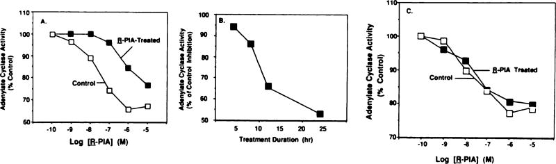
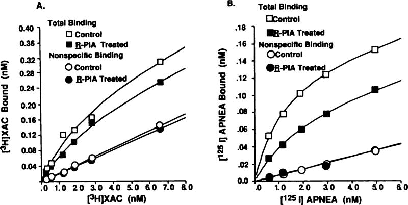
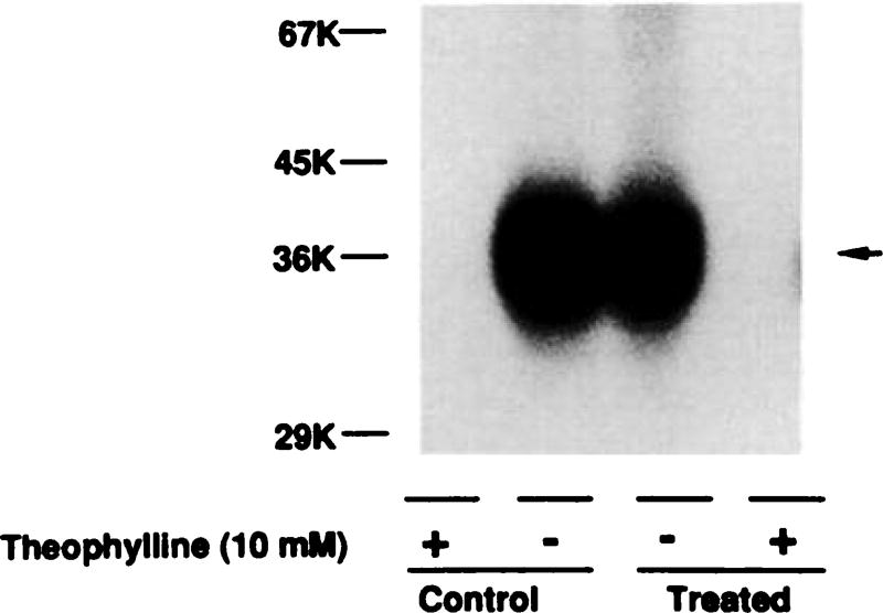
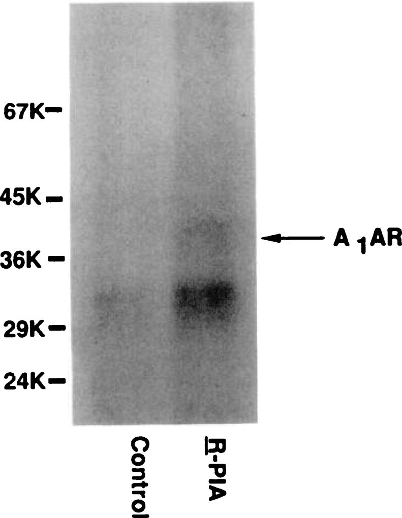
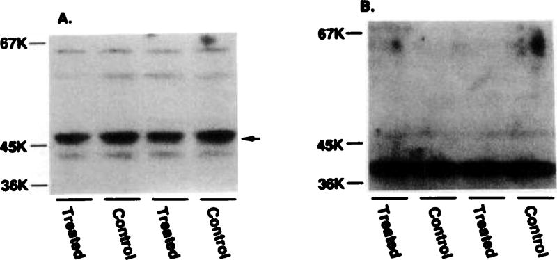
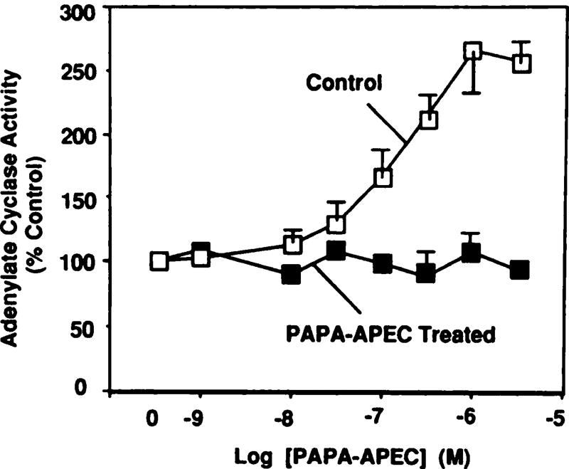
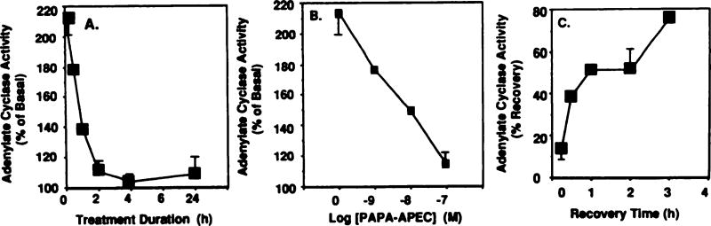
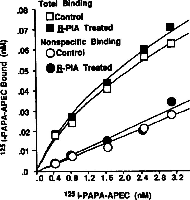
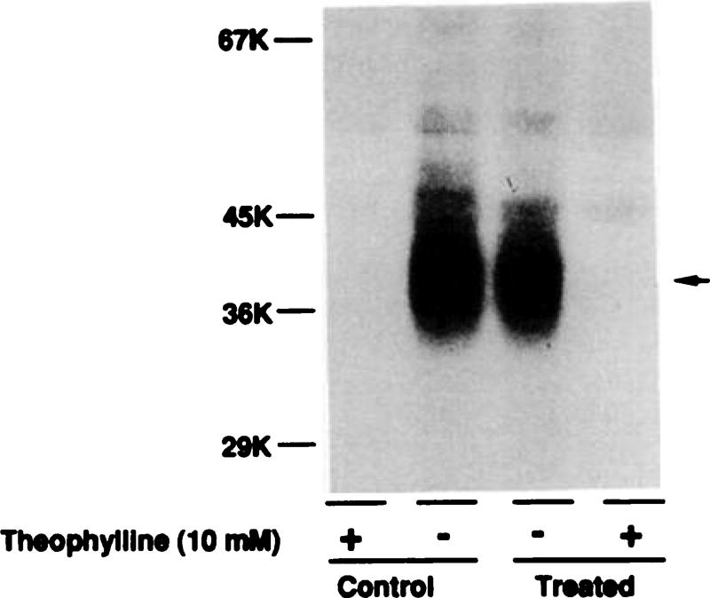
Desensitization of adenosine receptors (ARs) was studied in DDT1 MF-2 cells, which possess both A1- and A2AR, differentially coupled to adenylate cyclase. (-)-N6-(R)-Phenylisopropyladenosine (R-PIA), an A1AR-selective agonist at the appropriate concentrations, desensitized A1AR-mediated inhibition of adenylate cyclase activity in a time- (t1/2, 8 hr) and dose-dependent and reversible fashion. This was associated with significant decreases in total A1AR number and in the number of receptors possessing a high affinity for agonist in membrane preparations. The decrease in total A1AR in the membranes from the desensitized cells (approximately 40%) was associated with a 37% increase in A1AR measured in light vesicle preparations, compared with control cells. To test a possible role of phosphorylation in A1AR desensitization, cells were incubated with [32P]orthophosphate, followed by exposure to R-PIA for 18 hr. Subsequent purification of the A1AR indicated a 3-4-fold increase in phosphorylation of A1AR in cells treated with R-PIA, compared with control cells. Desensitization of the A1AR did not alter the levels of alpha s and alpha 12 proteins or affect the ability of stimulatory effectors, such as isoproterenol, sodium fluoride, and forskolin, to activate adenylate cyclase. These results suggest that uncoupling, down-regulation, and phosphorylation of the A1AR contribute, at least in part, to desensitization of this inhibitory receptor. Desensitization of the A2AR was characterized using an A2-selective agonist, 2-[4-(2-(4-aminophenyl]methylcarbonyl)ethyl)phenyl]ethylamino- 5'-N-ethylcarboxamidoadenosine (PAPA-APEC). Pretreatment of cells with PAPA-APEC (100 nM) resulted in a rapid loss of agonist stimulation of adenylate cyclase activity (t1/2 of this effect, 45 min). This effect was dose dependent (EC50, approximately 10 nM) and rapidly reversible. Interestingly, desensitization of the A2AR resulted in no change in receptor number, affinity, or mobility on sodium dodecyl sulfate-polyacrylamide gel electrophoresis. Taken together, these data suggest distinct mechanisms of desensitization of A1- and A2ARs in a single cell type.


Figures

Fig. 1
Fig. 1
Desensitization of A1AR-mediated inhibition of adenylate cyclase activity. A, DDT1 MF-2 cells were treated without (control) or with R-PIA (1 µm) for 24 hr, in the presence of adenosine deaminase. Adenylate cyclase assays were performed as described in Experimental Procedures. This experiment is representative of five experiments showing similar results. B, Time course of desensitization of A1AR. Inhibition of adenylate cyclase activity produced by R-PIA (1 µm) in control versus treated cells was determined at various times during drug treatment. Data are presented as the mean ± standard error of three independent experiments at each time point. C, Full R-PIA inhibition curves in control cells and those treated for 4 hr. Data presented are the mean of two experiments.
Fig. 2
Fig. 2
Quantitation of A1ARs in membranes prepared from control and R-PIA (1 µm, 24 hr)-treated cells, using the selective antagonist radioligand [3H]XAC (A) or selective agonist radioligand [125I]APNEA (B). Saturation curves were fitted by computer modeling, according to a one-state fit (19, 20). These curves are representative of five (A) or three (B) independent experiments.
Fig. 3
Fig. 3
Photoaffinity labeling of A1AR using [125I]AZPNEA. Membranes from control cells and cells treated with R-PIA (1 µm) for 24 hr were photolabeled with [125I]AZPNEA and subjected to SDS-PAGE, as described in Experimental Procedures. In order to test whether the migration of A1AR from treated cells was altered, a 50% increase in membrane proteins was added to the treated lanes. This is a representative figure of three independent experiments. After densitometric scanning of the autoradiograph and normalization for the different amounts of protein loaded per lane, incorporation of [125I]AZPNEA in the treated membranes was 40% of control. The average incorporation of [125I]AZPNEA into the desensitized membranes was 30 ± 9% of control (three experiments). Molecular weights of iodinated standards are indicated on the left.
Fig. 4
Fig. 4
Phosphorylation of A1AR in DDT1 MF-2 cells after R-PIA treatment. DDT1 MF-2 cells were incubated with [32P]orthophosphate for 2 hr at 37°, in phosphate-free DMEM. Cells were washed twice and then finally resuspended in DMEM containing 5% fetal bovine serum and 5% calf serum, with or without R-PIA (1 µm). After agonist treatment for 18 hr, cells were solubilized and A1ARs were purified by XAC-Affigel chromatography, followed by hydroxylapatite chromatography. The purified A1AR was resolved on 10% SDS-PAGE. Approximately 70 fmol of A1AR was loaded in the control lane and 40 fmol of A1AR in the R-PIA-treated lane.
Fig. 5
Fig. 5
Quantitation of αi and αs proteins by Western blotting in control and R-PIA-treated cell membranes. A, Membranes (~60 µg/lane) were solubilized and resolved on 10% SDS-polyacrylamide gels, as described previously (9, 19). Proteins were then transferred to nitrocellulose filters and were probed with antibody AS/7 (1/500 dilution), followed by 125IProtein A (300,000 cpm/ml). B, Filters were probed with the αs-specific antibody RM/1 (1/500 dilution).
Fig. 6
Fig. 6
Desensitization of A2AR-mediated activation of adenylate cyclase. Cells were treated with vehicle (control) or with PAPA-APEC (100 nm) for 24 hr, in the presence of adenosine deaminase (0.3 units/ml). Adenylate cyclase activity was assayed as described in Experimental Procedures. Basal activity in control and PAPA-APEC-treated cells averaged 2.2 ± 0.6 and 2.9 ± 1.3 pmol/min/mg of protein, respectively. Data are presented as the mean ± standard error of five independent experiments.
Fig. 7
Fig. 7
Desensitization of A2ARs by PAPA-APEC. A, DDT1 MF-2 smooth muscle cells were treated for various time periods with PAPA-APEC (100 nm). Basal adenylate cyclase activity averaged 2.0 pmol/min/mg of protein. PAPA-APEC (1 µm) was used to assess the extent of desensitization. B, Dose dependency of PAPA-APEC-mediated desensitization of activation of adenylate cyclase. Cells were treated with different concentrations of PAPA-APEC (1–100 nm) for 4 hr. Each point represents the mean ± standard error of two to four experiments. C, Time course of recovery from desensitization. Cells were treated with PAPA-APEC (100 nm) for 3 hr, after which they were washed and resuspended in fresh DMEM containing 5% serum and 0.3 units/ml adenosine deaminase. Data presented are the mean ± standard error of two or three independent experiments.
Fig. 8
Fig. 8
Quantitation of A2ARs using 125I-PAPA-APEC, in membranes from control and PAPA-APEC-treated cells. Cells were treated with PAPA-APEC (100 nm) for 24 hr at 37°. Saturation curves were performed using 125I-PAPA-APEC, as described in Experimental Procedures. Curves were fitted by a computer-assisted program (23, 24), assuming a one-state model. This is representative of five independent experiments showing similar results.
Fig. 9
Fig. 9
Photoaffinity labeling of A2AR in membranes prepared from control cells and cells treated with PAPA-APEC (100 nm) for 24 hr. Membranes were labeled with the A2AR-selective photoaffinity probe 125I-azidoPAPA-APEC (0.8 nm), as described in Experimental Procedures. Lanes 1 and 2, control membranes in the presence (lane 1) and absence (lane 2) of theophylline (10 mm) during the binding experiment; lanes 3 and 4, treated membranes in the absence (lane 3) and presence (lane 4) of theophylline. Specific 125I-azidoPAPA-APEC counts incorporated in lanes 2 and 3 were 1021 cpm and 1018 cpm, respectively. This experiment was repeated twice with similar results.

Similar articles

Cited by

References

    1. Van Calker D, Muller M, Hamprecht B. Adenosine regulates via two different receptors the accumulation of cyclic AMP in cultured brain cells. J. Neurochem. 1979;33:999–1005. - PubMed
    1. Stiles GL. Adenosine receptors and beyond: molecular mechanisms of physiological regulation. Clin. Res. 1990;38:10–18. - PubMed
    1. Sperelakis N. Regulation of calcium slow channels of cardiac and smooth muscles by adenine nucleotides. In: Pelleg A, Michelson EL, Freifus LS, editors. Cardiac Electrophysiology and Pharmacology of Adenosine and ATP: Basic and Clinical Aspects. Alan R. Liss, Inc.; New York: 1987. pp. 135–193. - PubMed
    1. Kurachi Y, Nakajima T, Sugimoto T. On the mechanism of activation of muscarinic K+ channels by adenosine in isolated atrial cells: involvement of GTP-binding proteins. Pfluegers Arch. 1986;407:264–274. - PubMed
    1. DeMazancourt P, Guidicelli Y. Guanine nucleotides and adenosine “Ri”-site analogues stimulate the membrane bound low Km cyclic AMP phosphodiesterase of rat adipocytes. FEBS Lett. 1984;173:385–388. - PubMed

Publication types

Substances

LinkOut - more resources