

The mouse fibula as a suitable bone for the study of functional adaptation to mechanical loading
- PMID: 19442626
- PMCID: PMC2671587
- DOI: 10.1016/j.bone.2008.12.026
The mouse fibula as a suitable bone for the study of functional adaptation to mechanical loading
Abstract
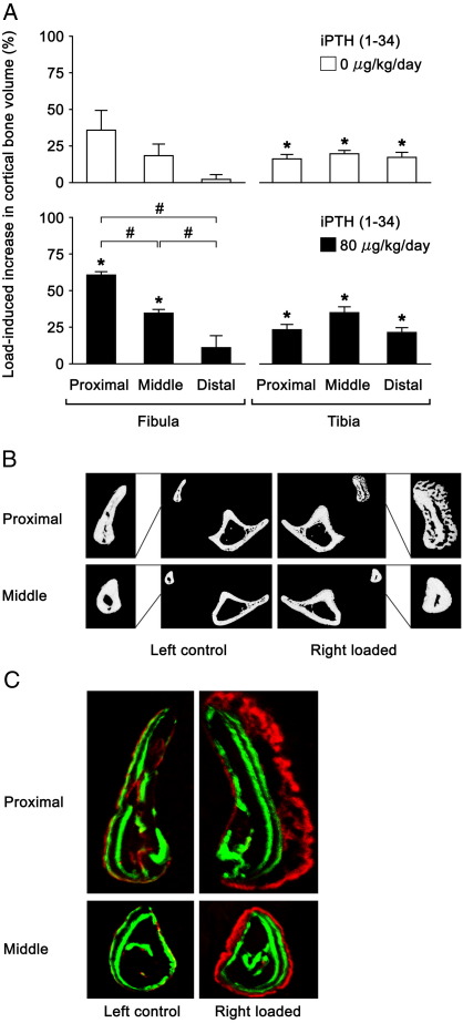
Bones' functionally adaptive responses to mechanical loading can usefully be studied in the tibia by the application of loads between the knee and ankle in normal and genetically modified mice. Such loading also deforms the fibula. Our present study was designed to ascertain whether the fibula adapts to loading in a similar way to the tibia and could thus provide an additional bone in which to study functional adaptation. The right tibiae/fibulae in C57BL/6 mice were subjected to a single period of axial loading (40 cycles at 10 Hz with 10-second intervals between each cycle; approximately 7 min/day, 3 alternate days/week, 2 weeks). The left tibiae/fibulae were used as non-loaded, internal controls. Both left and right fibulae and tibiae were analyzed by micro-computed tomography at the levels of the mid-shaft of the fibula and 25% from its proximal and distal ends. We also investigated the effects of intermittent parathyroid hormone (iPTH) on the (re)modelling response to 2-weeks of loading and the effect of 2-consecutive days of loading on osteocytes' sclerostin expression. These in vivo experiments confirmed that the fibula showed similar loading-related (re)modelling responses to those previously documented in the tibia and similar synergistic increases in osteogenesis between loading and iPTH. The numbers of sclerostin-positive osteocytes at the proximal and middle fibulae were markedly decreased by loading. Collectively, these data suggest that the mouse fibula, as well as the tibia and ulna, is a useful bone in which to assess bone cells' early responses to mechanical loading and the adaptive (re)modelling that this engenders.
Figures


References
-
- Ehrlich P.J., Lanyon L.E. Mechanical strain and bone cell function: a review. Osteoporos. Int. 2002;13:688–700. - PubMed
-
- O'Connor J.A., Lanyon L.E., MacFie H. The influence of strain rate on adaptive bone remodelling. J. Biomech. 1982;15:767–781. - PubMed
-
- Lanyon L.E., Rubin C.T. Static vs dynamic loads as an influence on bone remodelling. J. Biomech. 1984;17:897–905. - PubMed
-
- Pead M.J., Suswillo R., Skerry T.M., Vedi S., Lanyon L.E. Increased 3H-uridine levels in osteocytes following a single short period of dynamic bone loading in vivo. Calcif. Tissue Int. 1988;43:92–96. - PubMed
-
- Turner C.H., Akhter M.P., Raab D.M., Kimmel D.B., Recker R.R. A noninvasive, in vivo model for studying strain adaptive bone modeling. Bone. 1991;12:73–79. - PubMed
Publication types
MeSH terms
Substances
Grants and funding
LinkOut - more resources
Full Text Sources
