. 2009 Aug;35(2):219-33.
doi: 10.1016/j.nbd.2009.05.001. Epub 2009 May 12.

Dendritic spine pathologies in hippocampal pyramidal neurons from Rett syndrome brain and after expression of Rett-associated MECP2 mutations

Affiliations

Dendritic spine pathologies in hippocampal pyramidal neurons from Rett syndrome brain and after expression of Rett-associated MECP2 mutations

Christopher A Chapleau et al. Neurobiol Dis. 2009 Aug.

Abstract

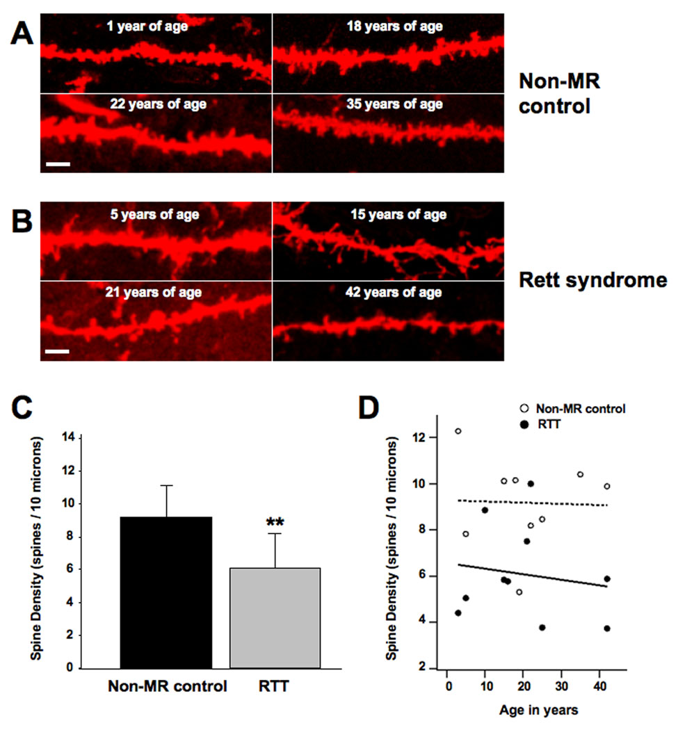
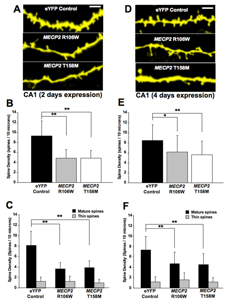
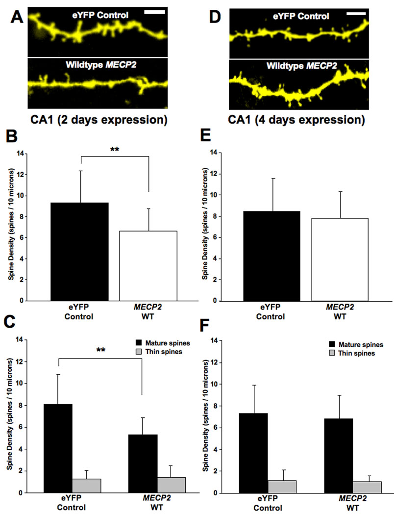
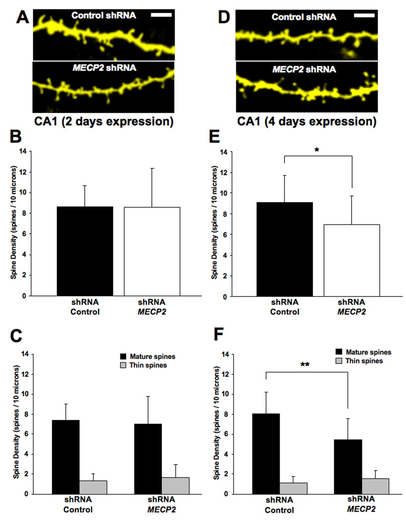
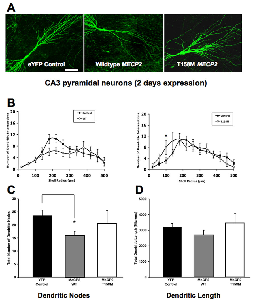
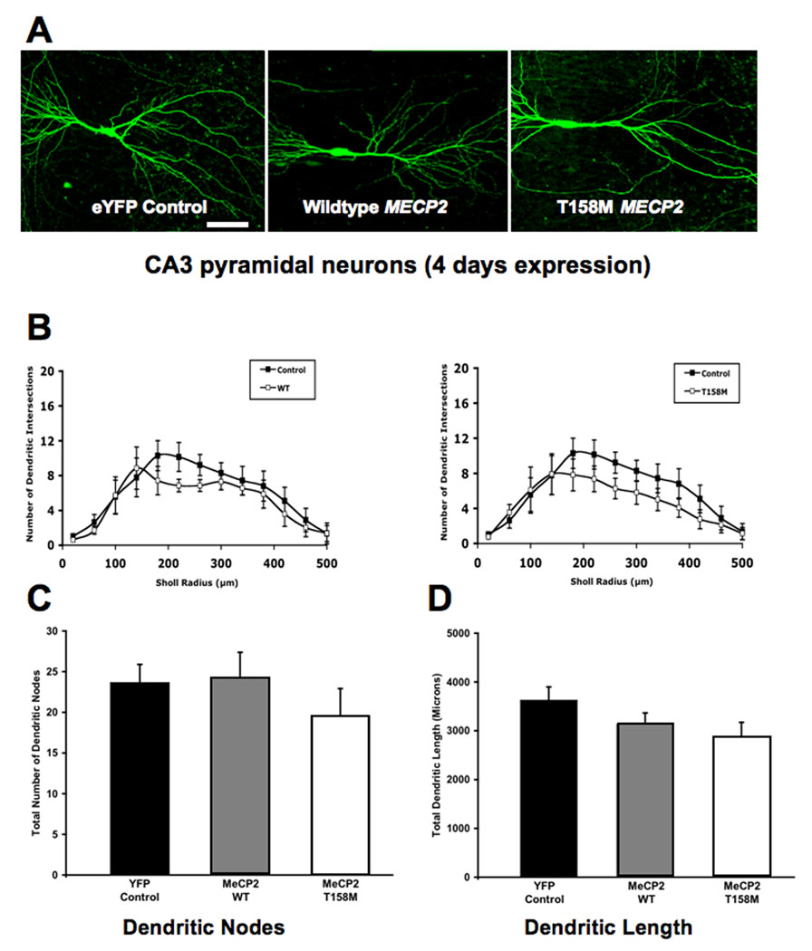
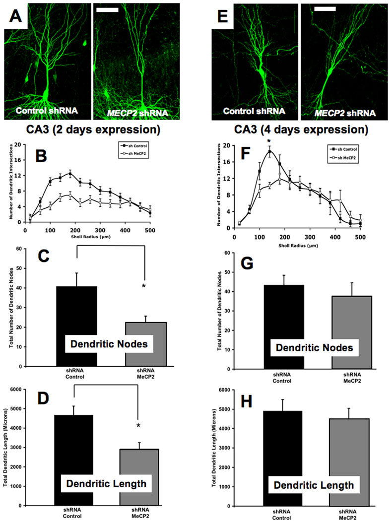
Rett syndrome (RTT) is an X chromosome-linked neurodevelopmental disorder associated with the characteristic neuropathology of dendritic spines common in diseases presenting with mental retardation (MR). Here, we present the first quantitative analyses of dendritic spine density in postmortem brain tissue from female RTT individuals, which revealed that hippocampal CA1 pyramidal neurons have lower spine density than age-matched non-MR female control individuals. The majority of RTT individuals carry mutations in MECP2, the gene coding for a methylated DNA-binding transcriptional regulator. While altered synaptic transmission and plasticity has been demonstrated in Mecp2-deficient mouse models of RTT, observations regarding dendritic spine density and morphology have produced varied results. We investigated the consequences of MeCP2 dysfunction on dendritic spine structure by overexpressing ( approximately twofold) MeCP2-GFP constructs encoding either the wildtype (WT) protein, or missense mutations commonly found in RTT individuals. Pyramidal neurons within hippocampal slice cultures transfected with either WT or mutant MECP2 (either R106W or T158M) showed a significant reduction in total spine density after 48 h of expression. Interestingly, spine density in neurons expressing WT MECP2 for 96 h was comparable to that in control neurons, while neurons expressing mutant MECP2 continued to have lower spine density than controls after 96 h of expression. Knockdown of endogenous Mecp2 with a specific small hairpin interference RNA (shRNA) also reduced dendritic spine density, but only after 96 h of expression. On the other hand, the consequences of manipulating MeCP2 levels for dendritic complexity in CA3 pyramidal neurons were only minor. Together, these results demonstrate reduced dendritic spine density in hippocampal pyramidal neurons from RTT patients, a distinct dendritic phenotype also found in neurons expressing RTT-associated MECP2 mutations or after shRNA-mediated endogenous Mecp2 knockdown, suggesting that this phenotype represent a cell-autonomous consequence of MeCP2 dysfunction.


Figures

FIGURE 1
FIGURE 1. QUANTITATIVE ANALYSIS IN HUMAN POSTMORTEM BRAIN HIPPOCAMPUS REVEALS THAT CA1 PYRAMIDAL NEURONS OF RTT INDIVIDUALS HAVE LOWER DENDRITIC SPINE DENSITY THAN THOSE OF NON-MR INDIVIDUALS
Formalin-fixed human brain samples were obtained from the Harvard Brain Bank and from the University of Maryland Brain Bank (Table 1 contains all available information on the individuals whose brains were used in this study). A. Representative examples of DiI-stained secondary and tertiary apical dendritic segments of CA1 pyramidal neurons from unaffected (non-MR) female individuals who served as controls. B. Representative examples of DiI-stained secondary and tertiary apical dendritic segments of CA1 pyramidal neurons from female RTT individuals. C. Dendritic spine density expressed per 10µm of dendritic length. D. Least square regression analyses of correlation between age and spine density in controls and RTT neurons. In this and all subsequent figures, data are presented as mean±SD, and * indicates p<0.05, ** indicates p<0.01, and *** indicates p<0.001, after unpaired two-tailed Student’s t test (see text for details). In this and all subsequent figures, scale bar represents 2µm.
FIGURE 2
FIGURE 2. OVEREXPRESSION OF RTT-ASSOCIATED MECP2 MUTATIONS CAUSES A PERSISTENT REDUCTION IN DENDRITIC SPINE DENSITY IN HIPPOCAMPAL PYRAMIDAL NEURONS
A. Representative examples of apical secondary or tertiary dendritic segments of CA1 pyramidal neurons expressing either an eYFP control plasmid or expression plasmids encoding GFP-tagged mutations in the methyl-binding domain of the MeCP2 (R106W or T158M) after 2 days of expression. B. Dendritic spine density expressed per 10µm of dendritic length. C. Density of each morphological type of dendritic spine (see text for classification criterion). D. Representative examples of apical secondary or tertiary dendritic segments of CA1 pyramidal neurons expressing either the eYFP control plasmid or mutant MECP2 after 4 days of expression). E. Dendritic spine density expressed per 10µm of dendritic length. F. Density of each morphological type of dendritic spine.
FIGURE 3
FIGURE 3. OVEREXPRESSION OF WILDTYPE MECP2 CAUSES A TRANSIENT REDUCTION IN DENDRITIC SPINE DENSITY IN HIPPOCAMPAL PYRAMIDAL NEURONS
A. Representative examples of apical secondary or tertiary dendritic segments of pyramidal neurons expressing either the eYFP control plasmid or wildtype MECP2 after 2 days of expression. B. Dendritic spine density expressed per 10µm of dendritic length. C. Density of each morphological type of dendritic spine. D. Representative examples of apical secondary or tertiary dendritic segments of CA1 pyramidal neurons expressing either the eYFP control plasmid or wildtype MECP2 after 4 days of expression. E. Dendritic spine density expressed per 10µm of dendritic length. F. Density of each morphological type of dendritic spine.
FIGURE 4
FIGURE 4. KNOCKDOWN OF ENDOGENOUS MECP2 CAUSES A REDUCTION IN THE DENSITY OF MATURE DENDRITIC SPINES ONLY AFTER 4 DAYS OF TRANSFECTION
A. Representative examples of apical secondary or tertiary dendritic segments of CA1 pyramidal neurons expressing either the eYFP control plasmid or an shRNA interfering sequence to knockdown endogenous Mecp2 after 2 days of expression. B. Dendritic spine density expressed per 10µm of dendritic length. C. Density of each morphological type of dendritic spine. D. Representative examples of apical secondary or tertiary dendritic segments of CA1 pyramidal neurons expressing either the eYFP control plasmid or an shRNA interfering sequence to knockdown endogenous Mecp2 after 4 days of expression. E. Dendritic spine density expressed per 10µm of dendritic length. F. Density of each morphological type of dendritic spine.
FIGURE 5
FIGURE 5. OVEREXPRESSION OF WILDTYPE MECP2 TRANSIENTLY REDUCES THE NUMBER OF DENDRITIC NODES AND THE TOTAL DENDRITIC LENGTH, WHILE T158M MECP2 TRANSIENTLY INCREASES DENDRITIC COMPLEXITY (2 DAYS OF EXPRESSION)
A. Representative low magnification views of eYFP-expressing CA3 pyramidal neurons transfected with either wildtype MECP2 or the T158M MECP2 mutant (2 days of expression). B. Three-dimensional Sholl analysis of dendritic complexity. Total number of intersections of CA3 apical dendrites as a function of distance from the soma. C. Total number of apical dendritic nodes. D. Total dendritic length. In this and Figure 6 and Figure 7, the scale bar represents 50µm.
FIGURE 6
FIGURE 6. OVEREXPRESSION OF WILDTYPE MECP2 TRANSIENTLY REDUCES THE NUMBER OF DENDRITIC NODES AND THE TOTAL DENDRITIC LENGTH, WHILE T158M MECP2 TRANSIENTLY INCREASES DENDRITIC COMPLEXITY (4 DAYS OF EXPRESSION)
A. Representative low magnification views of eYFP-expressing CA3 pyramidal neurons transfected with either wildtype MECP2 or the T158M MECP2 mutant (4 days of expression). B. Three-dimensional Sholl analysis of dendritic complexity. Total number of intersections of CA3 apical dendrites as a function of distance from the soma. C. Total number of apical dendritic nodes. D. Total dendritic length.
FIGURE 7
FIGURE 7. KNOCKDOWN OF ENDOGENOUS MECP2 DECREASES THE NUMBER OF DENDRITIC NODES, TOTAL DENDRITIC LENGTH, AND DENDRITIC INTERSECTIONS
A. Representative low magnification views of eYFP-expressing CA3 pyramidal neurons transfected with an shRNA control plasmid or a specific shRNA interfering sequence designed to knockdown endogenous Mecp2 (2 days of expression). B. Three-dimensional Sholl analysis of dendritic complexity. Total number of intersections of CA3 apical dendrites as a function of distance from the soma. C. Total number of apical dendritic nodes. D. Total dendritic length. E. Representative low magnification views of eYFP-expressing CA3 pyramidal neurons transfected with the shRNA control plasmid or the specific Mecp2 shRNA (4 days of expression). F. Three-dimensional Sholl analysis of dendritic complexity. Total number of intersections of CA3 apical dendrites as a function of distance from the soma. G. Total number of apical dendritic nodes. H. Total dendritic length.

References

    1. Akbarian S, Chen RZ, Gribnau J, Rasmussen TP, Fong H, Jaenisch R, Jones EG. Expression pattern of the Rett syndrome gene MeCP2 in primate prefrontal cortex. Neurobiol Dis. 2001;8:784–791. - PubMed
    1. Alonso M, Medina JH, Pozzo-Miller L. ERK1/2 activation is necessary for BDNF to increase dendritic spine density in hippocampal CA1 pyramidal neurons. Learn Mem. 2004;11:172–178. - PMC - PubMed
    1. Amir RE, Van den Veyver IB, Wan M, Tran CQ, Francke U, Zoghbi HY. Rett syndrome is caused by mutations in X-linked MECP2, encoding methyl-CpG-binding protein 2. Nat Genet. 1999;23:185–188. - PubMed
    1. Armstrong D, Dunn JK, Antalffy B, Trivedi R. Selective dendritic alterations in the cortex of Rett syndrome. J Neuropathol Exp Neurol. 1995;54:195–201. - PubMed
    1. Asaka Y, Jugloff DG, Zhang L, Eubanks JH, Fitzsimonds RM. Hippocampal synaptic plasticity is impaired in the Mecp2-null mouse model of Rett syndrome. Neurobiol Dis. 2006;21:217–227. - PubMed

Publication types

MeSH terms