. 2009 May;84(5):706-11.
doi: 10.1016/j.ajhg.2009.04.016.

DYNC2H1 mutations cause asphyxiating thoracic dystrophy and short rib-polydactyly syndrome, type III

Affiliations

DYNC2H1 mutations cause asphyxiating thoracic dystrophy and short rib-polydactyly syndrome, type III

Nathalie Dagoneau et al. Am J Hum Genet. 2009 May.

Abstract

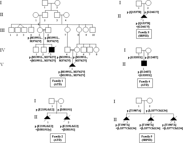
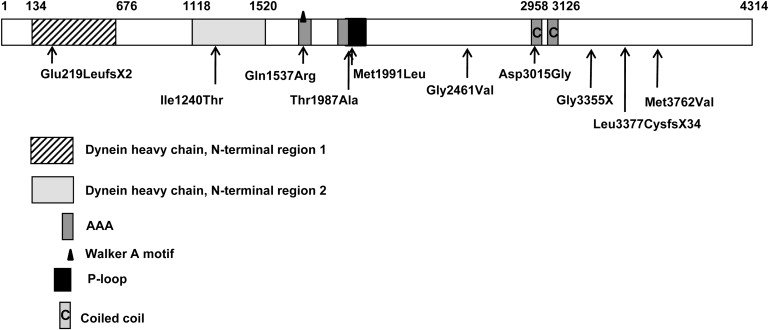
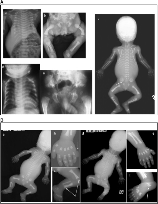
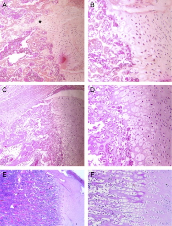
Jeune asphyxiating thoracic dystrophy (ATD) is an autosomal-recessive chondrodysplasia characterized by short ribs and a narrow thorax, short long bones, inconstant polydactyly, and trident acetabular roof. ATD is closely related to the short rib polydactyly syndrome (SRP) type III, which is a more severe condition characterized by early prenatal expression and lethality and variable malformations. We first excluded IFT80 in a series of 26 fetuses and children belonging to 14 families diagnosed with either ATD or SRP type III. Studying a consanguineous family from Morocco, we mapped an ATD gene to chromosome 11q14.3-q23.1 in a 20.4 Mb region and identified homozygous mutations in the cytoplasmic dynein 2 heavy chain 1 (DYNC2H1) gene in the affected children. Compound heterozygosity for DYNC2H1 mutations was also identified in four additional families. Among the five families, 3/5 were diagnosed with ATD and 2/5 included pregnancies terminated for SRP type III. DYNC2H1 is a component of a cytoplasmic dynein complex and is directly involved in the generation and maintenance of cilia. From this study, we conclude that ATD and SRP type III are variants of a single disorder belonging to the ciliopathy group.


Figures

Figure 1
Figure 1
Pedigrees of the Five Affected Families and Segregation of the DYNC2H1 Mutations
Figure 2
Figure 2
Location of the DYNC2H1 Mutations
Figure 3
Figure 3
Tridimensional Structure Model of a DYNC2H1 Putative Microtubule-Binding Domain Via Swiss-Pdb Viewer 3.7 Representation The modeled protein is represented in orange and is superimposed with the template (PDB code: 3err) shown in gray. Between Asp3015 (in red) and Asn3070 (in green), a putative hydrogen may be computed. The mutation p.Asp3015Gly disrupts this stabilizing hydrogen bond, inducing a local conformational change altering the function of this putative microtubule-binding domain.
Figure 4
Figure 4
Radiological Findings in the Families with DYNC2H1 Mutations (A) ATD cases. a and b: case 1 (day 2) from family 1; c: case 1 (28 wg) from family 2; d and e: affected child (4 mo) from family 4. Note the short bones, narrow thorax, and trident aspect of the acetabular roof (c, arrow). Note also the advanced proximal femoral ossification in the family 4 case (e). (B) SRP cases. a–c: terminated pregnancy at 25 wg from family 3; d–f: case 1 from family 5. Note the severe shortness of the long bones, the narrow thorax, and the trident aspect of the acetabular roof. Note also the round metaphyseal ends with lateral spikes (c, arrows), the triplicate calcaneum (d, arrow), and the postaxial polydactyly (a, arrow). Note the bowing of the femora observed in the cases from families 1–3 and 5 (Ab, Ac, Bc, Bd).
Figure 5
Figure 5
Femoral Histology in the Two SRP Type III Patients and in a Control Fetus Physeal growth zone shows short and irregular columns in the proliferative zone in the family 5 case (A×10, B×20) and in the family 3 case (C×10, D×20), compared to a control of the same gestational age (E×10, F×20). Note also the more severe disorganization in the family 5 case, with associated elongated tongues of hypertrophic cartilage (asterisks) and patchy cellular proliferation.

References

    1. Beales P.L., Bland E., Tobin J.L., Bacchelli C., Tuysuz B., Hill J., Rix S., Pearson C.G., Kai M., Hartley J. IFT80, which encodes a conserved intraflagellar transport protein, is mutated in Jeune asphyxiating thoracic dystrophy. Nat. Genet. 2007;39:727–729. - PubMed
    1. Morgan N.V., Bacchelli C., Gissen P., Morton J., Ferrero G.B., Silengo M., Labrune P., Casteels I., Hall C., Cox P. A locus for asphyxiating thoracic dystrophy, ATD, maps to chromosome 15q13. J. Med. Genet. 2003;40:431–435. - PMC - PubMed
    1. Superti-Furga A., Unger S. Nosology and classification of genetic skeletal disorders: 2006 revision. Am. J. Med. Genet. 2007;143:1–18. - PubMed
    1. Ho N.C., Francomano C.A., van Allen M. Jeune asphyxiating thoracic dystrophy and short-rib polydactyly type III (Verma-Naumoff) are variants of the same disorder. Am. J. Med. Genet. 2000;90:310–314. - PubMed
    1. Pfister K.K., Shah P.R., Hummerich H., Russ A., Cotton J., Annuar A.A., King S.M., Fisher E.M. Genetic analysis of the cytoplasmic dynein subunit families. PLoS Genet. 2006;2:e1. - PMC - PubMed

Publication types

LinkOut - more resources