

Generation and characterization of JCV permissive hybrid cell lines
- PMID: 19442856
- PMCID: PMC2692597
- DOI: 10.1016/j.jviromet.2009.02.023
Generation and characterization of JCV permissive hybrid cell lines
Abstract
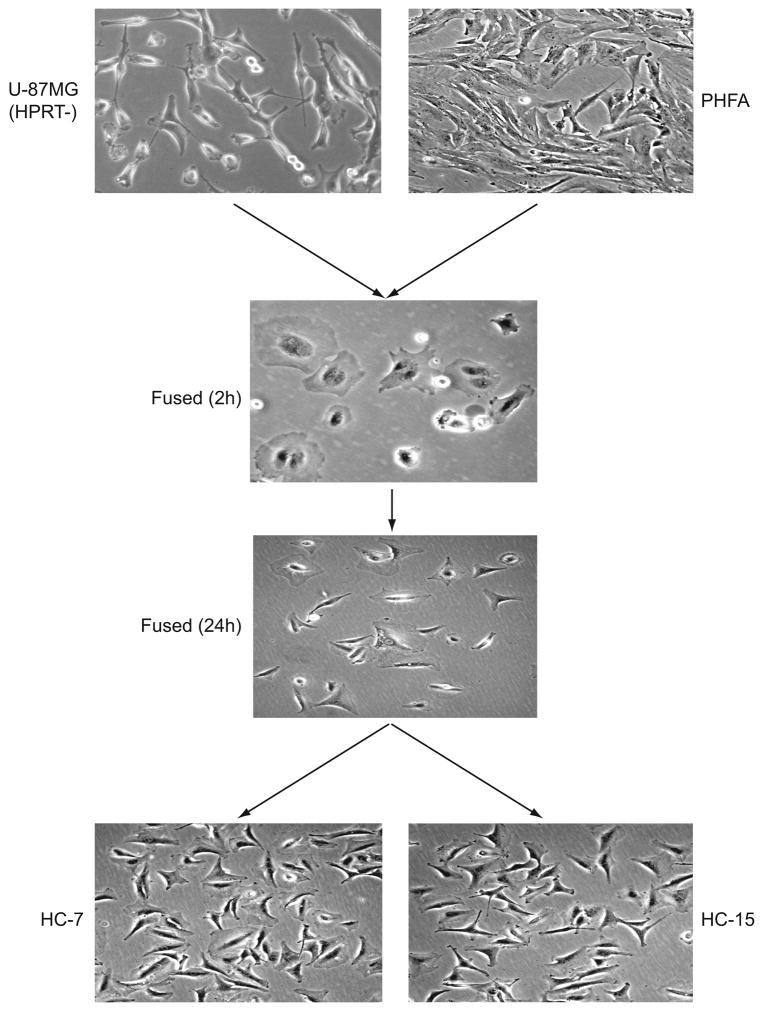
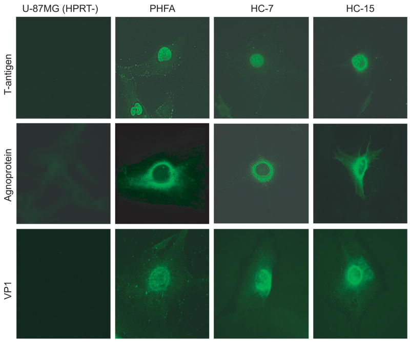
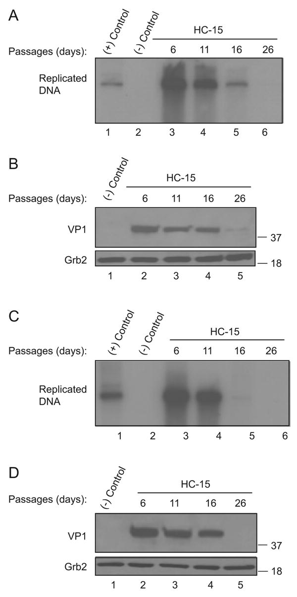
JC virus (JCV) is a human neurotropic polyomavirus whose replication in the central nervous system induces the fatal demyelinating disease, progressive multifocal leukoencephalopathy (PML). JCV particles have been detected primarily in oligodendrocytes and astrocytes of the brains of patients with PML and in the laboratory its propagation is limited to primary cultures of human fetal glial cells. In this short communication, the development of a new cell culture system is described through the fusion of primary human fetal astrocytes with the human glioblastoma cell line, U-87MG. The new hybrid cell line obtained from this fusion has the capacity to support efficiently expression of JCV and replication of viral DNA in vitro up to 16 passages. This cell line can serve as a reliable culture system to study the biology of JCV host-cell interaction, determine the mechanisms involved in cell type specific replication of JCV, and provide a convenient cell culture system for high throughput screening of anti-viral agents.
Figures


Similar articles
-
Replication of JC Virus DNA in the G144 Oligodendrocyte Cell Line Is Dependent Upon Akt.J Virol. 2017 Sep 27;91(20):e00735-17. doi: 10.1128/JVI.00735-17. Print 2017 Oct 15. J Virol. 2017. PMID: 28768870 Free PMC article.
-
Deep-Sequence Identification and Role in Virus Replication of a JC Virus Quasispecies in Patients with Progressive Multifocal Leukoencephalopathy.J Virol. 2016 Dec 16;91(1):e01335-16. doi: 10.1128/JVI.01335-16. Print 2017 Jan 1. J Virol. 2016. PMID: 27795410 Free PMC article.
-
Human iPS cell-derived astrocytes support efficient replication of progressive multifocal leukoencephalopathy-type JC polyomavirus.Biochem Biophys Res Commun. 2020 Dec 17;533(4):983-987. doi: 10.1016/j.bbrc.2020.09.117. Epub 2020 Sep 30. Biochem Biophys Res Commun. 2020. PMID: 33008586
-
The Role of the JC Virus in Central Nervous System Tumorigenesis.Int J Mol Sci. 2020 Aug 28;21(17):6236. doi: 10.3390/ijms21176236. Int J Mol Sci. 2020. PMID: 32872288 Free PMC article. Review.
-
Traffic of JC virus from sites of initial infection to the brain: the path to progressive multifocal leukoencephalopathy.J Infect Dis. 2002 Dec 1;186 Suppl 2:S180-6. doi: 10.1086/344280. J Infect Dis. 2002. PMID: 12424695 Review.
Cited by
-
Clonal immortalized human glial cell lines support varying levels of JC virus infection due to differences in cellular gene expression.J Neuroimmune Pharmacol. 2013 Dec;8(5):1303-19. doi: 10.1007/s11481-013-9499-8. Epub 2013 Sep 20. J Neuroimmune Pharmacol. 2013. PMID: 24052414
-
The agnoprotein of polyomavirus JC is released by infected cells: evidence for its cellular uptake by uninfected neighboring cells.Virology. 2014 Nov;468-470:88-95. doi: 10.1016/j.virol.2014.07.054. Epub 2014 Aug 23. Virology. 2014. PMID: 25151063 Free PMC article.
-
Exploring the role of NCCR variation on JC polyomavirus expression from dual reporter minicircles.PLoS One. 2018 Jun 26;13(6):e0199171. doi: 10.1371/journal.pone.0199171. eCollection 2018. PLoS One. 2018. PMID: 29944671 Free PMC article.
-
Polyomavirus large T antigens: Unraveling a complex interactome.Tumour Virus Res. 2025 Jun;19:200306. doi: 10.1016/j.tvr.2024.200306. Epub 2024 Dec 13. Tumour Virus Res. 2025. PMID: 39675526 Free PMC article. Review.
-
Molecular biology, epidemiology, and pathogenesis of progressive multifocal leukoencephalopathy, the JC virus-induced demyelinating disease of the human brain.Clin Microbiol Rev. 2012 Jul;25(3):471-506. doi: 10.1128/CMR.05031-11. Clin Microbiol Rev. 2012. PMID: 22763635 Free PMC article. Review.
References
-
- Akan I, Sariyer IK, Biffi R, Palermo V, Woolridge S, White MK, Amini S, Khalili K, Safak M. Human polyomavirus JCV late leader peptide region contains important regulatory elements. Virology. 2006;349:66–78. - PubMed
-
- Akatani K, Imai M, Kimura M, Nagashima K, Ikegami N. Propagation of JC virus in human neuroblastoma cell line IMR-32. J Med Virol. 1994;43:13–19. - PubMed
-
- Amano T, Hamprechy B, Kemper W. High activity of choline acetyltransferase induced in neuroblastoma × glia hybrid cells. Exp Cell Res. 1974;85:399–408. - PubMed
-
- Ashok A, Atwood WJ. Polyomavirus receptors and tropism. Adv Exp Med Biol. 2006;577:60–72. - PubMed
Publication types
MeSH terms
Substances
Grants and funding
LinkOut - more resources
Full Text Sources
