

Review
doi: 10.1016/j.sbi.2009.04.002.
Epub 2009 May 13.
Structural insights into RNA splicing
Affiliations
- PMID: 19443210
- PMCID: PMC2756803
- DOI: 10.1016/j.sbi.2009.04.002
Item in Clipboard
Review
Structural insights into RNA splicing
Curr Opin Struct Biol.
2009 Jun.
Abstract
Intron splicing is a fundamental biological process whereby noncoding sequences are removed from precursor RNAs. Recent work has provided new insights into the structural features and reaction mechanisms of two introns that catalyze their own splicing from precursor RNA: the group I and II introns. In addition, there is an increasing amount of structural information on the spliceosome, which is a ribonucleoprotein machine that catalyzes nuclear pre-mRNA splicing in eukaryotes. Here, we compare structures and catalytic mechanisms of self-splicing RNAs and we discuss the possible implications for spliceosomal reaction mechanisms.
Figures
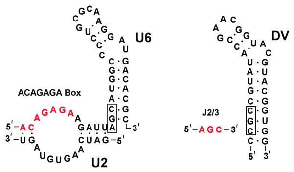
A comparison of the secondary structures of the U6 snRNA from the spliceosome (left) and domain V from the O. iheyensis group II intron (right). The catalytic triad (boxed) is located at the base of the stem loop structures, with the two-nucleotide bulge spaced five base pairs away. The ACAGGA box (red) may form base triple interactions with the triad of the U6 snRNA, analogous to that observed between J2/3 and domain V.

RNA secondary structures of the IIA, IIB, and IIC intron classes. The IIC class is smaller and more compact, especially in domain I. Tertiary interactions are indicated with Greek letters and domains with Roman numerals.

Overall tertiary structure of the group II intron. Domain V (red) forms the catalytic core with the rest of the domains encapsulating this active site.

Comparison of the group I and group II active sites with bound exons. (a) In the group II intron, putatively catalytic metal ions (M1 and M2) are within inner shell coordination distance of the scissile phosphate at the exon junction. Note that other metal ions, omitted for clarity, have also been identified within the intron core (reference). (b) The group I structure represents a fully active intron with ~70% of the pre-ligated form (transparent) and ~30% of the ligated (solid). The scissile phosphate of the ligated exons is not within coordination distance (~4.5-6 Å) of the metal ions. In the pre-ligated state, the distance changes to ~2.5 Å, which is appropriate for coordination. The group II structure is analogous to the ligated, product form of the group I intron.
References
-
- Vicens Q, Cech T. Atomic level architecture of group I introns revealed. Trends Biochem Sci. 2006;31:41–51. - PubMed
-
- Guerrier-Takada C, Gardiner K, Marsh T, Pace N, Altman S. The RNA moiety of ribonuclease P is the catalytic subunit of the enzyme. Cell. 1983;35:849–857. - PubMed
-
- Peebles C, Perlman P, Mecklenburg K, Petrillo M, Tabor J, Jarrell K, Cheng H. A self-splicing RNA excises an intron lariat. Cell. 1986;44:213–223. - PubMed
-
- Pyle A, Fedorova O, Waldsich C. Folding of group II introns: a model system for large, multidomain RNAs? Trends Biochem Sci. 2007;32:138–145. - PubMed
-
- Michel F, Ferat J. Structure and activities of group II introns. Annu Rev Biochem. 1995;64:435–461. - PubMed
Publication types
MeSH terms
Substances
Grants and funding
LinkOut - more resources
Full Text Sources
Other Literature Sources
