. 2009 Jul;37(13):4264-74.
doi: 10.1093/nar/gkp390. Epub 2009 May 13.

ROS1 5-methylcytosine DNA glycosylase is a slow-turnover catalyst that initiates DNA demethylation in a distributive fashion

Affiliations

ROS1 5-methylcytosine DNA glycosylase is a slow-turnover catalyst that initiates DNA demethylation in a distributive fashion

María Isabel Ponferrada-Marín et al. Nucleic Acids Res. 2009 Jul.

Abstract

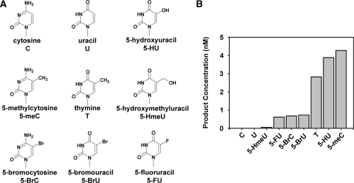
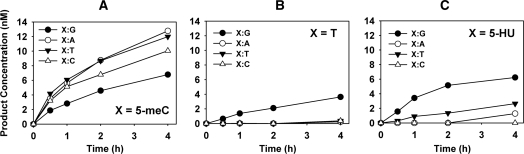
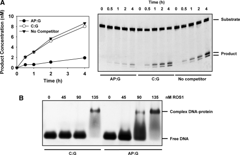
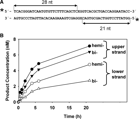
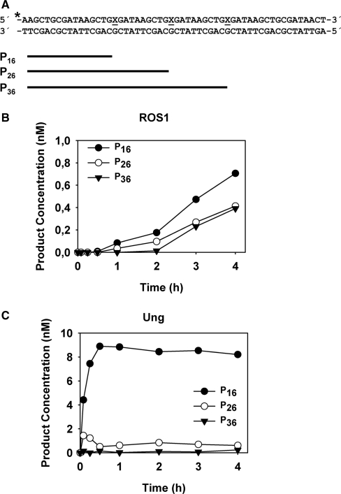
Arabidopsis ROS1 belongs to a family of plant 5-methycytosine DNA glycosylases that initiate DNA demethylation through base excision. ROS1 displays the remarkable capacity to excise 5-meC, and to a lesser extent T, while retaining the ability to discriminate effectively against C and U. We found that replacement of the C5-methyl group by halogen substituents greatly decreased excision of the target base. Furthermore, 5-meC was excised more efficiently from mismatches, whereas excision of T only occurred when mispaired with G. These results suggest that ROS1 specificity arises by a combination of selective recognition at the active site and thermodynamic stability of the target base. We also found that ROS1 is a low-turnover catalyst because it binds tightly to the abasic site left after 5-meC removal. This binding leads to a highly distributive behaviour of the enzyme on DNA substrates containing multiple 5-meC residues, and may help to avoid generation of double-strand breaks during processing of bimethylated CG dinucleotides. We conclude that the biochemical properties of ROS1 are consistent with its proposed role in protecting the plant genome from excess methylation.


Figures

Figure 1.
Figure 1.
ROS1 incision activity on duplex DNA substrates containing different modified bases paired with G. (A) Chemical structures of substrate DNA bases tested in this study. (B) Purified ROS1 (22.5 nM) was incubated at 30°C for 2 h with 51-mer double-stranded oligonucleotide substrates (20 nM) containing at position 29 of the labelled upper-strand different target DNA bases paired with G. Products were separated in a 12% denaturing polyacrylamide gel and the amount of incised oligonucleotide was quantified by fluorescent scanning.
Figure 2.
Figure 2.
Effect of base-pairing partner on ROS1 activity for 5-meC, T and 5-HU. The time-dependent generation of incision products was measured by incubating purified ROS1 (22.5 nM) at 30°C with double-stranded oligonucleotide substrates (20 nM) containing 5-meC (A), T (B) or 5-HU (C) opposite G (filled circles), A (open circles), T (filled triangles) or C (open triangles) on the complementary strand. Reactions were stopped at the indicated times, products were separated in a 12% denaturing polyacrylamide gel, and the amount of incised oligonucleotide was quantified by fluorescence scanning.
Figure 3.
Figure 3.
DNA glycosylase and AP lyase activities of ROS1. The time-dependent generation of incision products was measured by incubating purified ROS1 (22.5 nM) at 30°C with double-stranded oligonucleotide substrates (20 nM) containing a single 5-meC:G pair. Reactions were stopped at the indicated times and products were separated in a 12% denaturing polyacrylamide gel. Filled circles: nick incisions detected without alkaline treatment. Open circles: nick incisions detected after incubation with 100 mM NaOH at 90°C for 10 min, in order to cleave AP sites. The amount of incised oligonucleotide was quantified by fluorescence scanning.
Figure 4.
Figure 4.
Time-dependent product accumulation in reactions with different enzyme concentrations. (A) Different amounts of purified ROS1 (2.25 nM, filled circles; 4.5 nM, open circles; 22.5 nM, filled triangles), were incubated at 30°C with a double-stranded oligonucleotide substrate (20 nM) containing a 5-meC:G pair. (B and C) Reaction was started by addition of ROS1 (4.5 nM) and additional enzyme aliquots (4.5 nM) were added at 4 and 8 h (arrows). Reactions were stopped at the indicated times, products were separated in a 12% denaturing polyacrylamide gel (C), and the amount of incised oligonucleotide was quantified by fluorescence scanning (B).
Figure 5.
Figure 5.
ROS1 binding to DNA containing an AP site. (A) Effect of preincubation with DNA containing an AP site on ROS1 activity. Purified ROS1 (22.5 nM) was preincubated for 30 min with reaction buffer (filled triangles) or with 40 nM unlabelled duplex oligonucleotide containing either a synthetic AP site opposite guanine (AP:G, filled circles) or a C:G pair (open circles). Then, the fluorescein-labelled 5-meC:G substrate (40 nM) was added and the reactions were monitored for 4 h. Products were separated in a 12% denaturing polyacrylamide gel, and the relative amount of incised oligonucleotide was quantified by fluorescence scanning. (B) EMSA. Increasing amounts of ROS1 were incubated with a labelled duplex oligonucleotide containing either a synthetic AP site opposite guanine (AP:G) or a C:G pair. After nondenaturing gel electrophoresis, protein–DNA complexes were identified by their retarded mobility compared with that of free DNA, as indicated.
Figure 6.
Figure 6.
ROS1 activity on hemimethylated and bimethylated DNA. (A) Structure and length of the DNA substrates and products. X indicates target base. (B) Purified ROS1 (22.5 nM) was incubated with 20 nM duplex DNA labelled on both strands that contained a single hemimethylated or a bimethylated CG site. Reactions were stopped at the indicated times and products were separated in a 12% denaturing polyacrylamide gel. The number of incisions on the upper (filled symbols) and/or lower (open symbols) strand on the hemimethylated (circles) and bimethylated (triangle) DNA substrate was quantified by fluorescence scanning.
Figure 7.
Figure 7.
Processivity analysis of ROS1 activity. (A) Structure and length of the DNA substrates and products. X indicates target base. (B) Purified ROS1 (4.5 nM) was incubated with a double-stranded oligonucleotide substrate (10 nM) containing three 5-meC:G pairs. Reactions were stopped at the indicated times and products were separated in a 12% denaturing polyacrylamide gel. Product concentration was quantified by fluorescence scanning: 16 nt (filled circles), 26 nt (open circles) and 36 nt (filled triangles). (C) A control reaction was performed incubating purified E. coli Ung (1 × 10⁻³ U) with an equivalent double-stranded oligonucleotide substrate (10 nM) containing three U:G pairs.

Similar articles

Cited by

References

    1. Goll MG, Bestor TH. Eukaryotic cytosine methyltransferases. Annu. Rev. Biochem. 2005;74:481–514. - PubMed
    1. Bernstein BE, Meissner A, Lander ES. The mammalian epigenome. Cell. 2007;128:669–681. - PubMed
    1. Henderson IR, Jacobsen SE. Epigenetic inheritance in plants. Nature. 2007;447:418–424. - PubMed
    1. Bird A. DNA methylation patterns and epigenetic memory. Genes Dev. 2002;16:6–21. - PubMed
    1. Jones PA, Takai D. The role of DNA methylation in mammalian epigenetics. Science. 2001;293:1068–1070. - PubMed

Publication types