. 2009 Aug;155(Pt 8):2509-2521.
doi: 10.1099/mic.0.028852-0. Epub 2009 May 14.

Growth of calcium-blind mutants of Yersinia pestis at 37 degrees C in permissive Ca2+-deficient environments

Affiliations

Growth of calcium-blind mutants of Yersinia pestis at 37 degrees C in permissive Ca2+-deficient environments

Janet M Fowler et al. Microbiology (Reading). 2009 Aug.

Abstract

Cells of wild-type Yersinia pestis exhibit a low-calcium response (LCR) defined as bacteriostasis with expression of a pCD-encoded type III secretion system (T3SS) during cultivation at 37 degrees C without added Ca(2+) versus vegetative growth with downregulation of the T3SS with Ca(2+) (>or=2.5 mM). Bacteriostasis is known to reflect cumulative toxicity of Na(+), l-glutamic acid and culture pH; control of these variables enables full-scale growth ('rescue') in the absence of Ca(2+). Several T3SS regulatory proteins modulate the LCR, because their absence promotes a Ca(2+)-blind phenotype in which growth at 37 degrees C ceases and the T3SS is constitutive even with added Ca(2+). This study analysed the connection between the LCR and Ca(2+) by determining the response of selected Ca(2+)-blind mutants grown in Ca(2+)-deficient rescue media containing Na(+) plus l-glutamate (pH 5.5), where the T3SS is not expressed, l-glutamate alone (pH 6.5), where l-aspartate is fully catabolized, and Na(+) alone (pH 9.0), where the electrogenic sodium pump NADH : ubiquinone oxidoreductase becomes activated. All three conditions supported essentially full-scale Ca(2+)-independent growth at 37 degrees C of wild-type Y. pestis as well as lcrG and yopN mutants (possessing a complete but dysregulated T3SS), indicating that bacteriostasis reflects a Na(+)-dependent lesion in bioenergetics. In contrast, mutants lacking the negative regulator YopD or the YopD chaperone (LcrH) failed to grow in any rescue medium and are therefore truly temperature-sensitive. The Ca(2+)-blind yopD phenotype was fully suppressed in a Ca(2+)-independent background lacking the injectisome-associated inner-membrane component YscV but not peripheral YscK, suggesting that the core translocon energizes YopD.


Figures
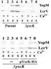
Fig. 1.
Fig. 1.
Complementation of the ΔyscK Y. pestis mutant used in this work. Non-polarity of the deletion within yscK in Y. pestis KIM8-3002.12 was tested by comparing the LCR phenotypes of the parental Y. pestis KIM8-3002, ΔyscK Y. pestis KIM8-3002.12 and the Y. pestis KIM8-3002.12 pYscKHA18 (YscK+) isolates. The bacteria were grown in TMH with (+) or without (−) 2.5 mM CaCl2 for 4 h at 37 °C, and YopM and LcrV abundance was assessed by immunoblotting in the bacterial cells (c) or the medium containing secreted proteins (s). Top panel, the yscK deletion mutant was compared for its Yops expression and secretion (lanes 5–8) with the parental strain (lanes 1–4); bottom panel, complementation by the haemagglutinin-tagged YscK (YscK-HA) expressed from pYscKHA18 was assessed by comparing Yops expression and secretion of the complemented strain (lanes 5–8) with those of the yscK mutant (lanes 1–4).
Fig. 2.
Fig. 2.
Growth of Lcr+ (•), Lcr (○), LcrG (▴), LcrV (▵), LcrH (▾), YopD (⧫) and YopN (▪) mutants of Y. pestis strain KIM in chemically defined medium containing 4.0 mM Ca2+ with (a) 100 mM Na+ and 25 mM l-glutamic acid (pH 7.5), (b) no added Na+ and 25 mM l-glutamic acid (pH 6.5), (c) 100 mM Na+ and no added l-glutamic acid (pH 9.0), and (d) 100 mM Na+ and 25 mM l-glutamic acid (pH 5.5). The dashed line indicates agglutination of yopD cells with precipitated calcium phosphate at pH 9.0.
Fig. 3.
Fig. 3.
Growth of Lcr+ (•), Lcr (○), LcrG (▴), LcrV (▵), LcrH (▾), YopD (⧫) and YopN (▪) mutants of Y. pestis strain KIM in chemically defined medium lacking added Ca2+ with (a) 100 mM Na+ and 25 mM l-glutamic acid (pH 7.5), (b) no added Na+ and 25 mM l-glutamic acid (pH 6.5), (c) 100 mM Na+ and no added l-glutamic acid (pH 9.0), and (d) 100 mM Na+ and 25 mM l-glutamic acid (pH 5.5).
Fig. 4.
Fig. 4.
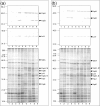
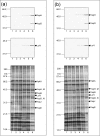
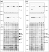
Immunoblots (YopH, YopD and LcrV) and silver-stained preparations following SDS-PAGE of whole cultures of Lcr, Pla (lane 1); Lcr+, Pla+ (lane 2); Lcr+, Pla (lane 3); lcrG, Pla (lane 4); lcrV, Pla (lane 5); lcrH, Pla (lane 6); yopD, Pla (lane 7); and yopN, Pla (lane 8) cells of Y. pestis strain KIM after 7 h incubation at 37 °C in chemically defined medium with 100 mM Na+ and 25 mM l-glutamic acid (pH 7.5) either containing (a) or lacking (b) added 4.0 mM Ca2+; also shown in both panels are Ca2+-deficient control cultures of Lcr+, Pla cells following growth for 9 h in the same Ca2+-deficient medium (lane 9). The amount of the ∼19 kDa band corresponding to fibrillar capsular antigen fraction 1 (Caf1) (open arrows) varied in amount among the strains and correlated with their net growth following shift from 26 to 37 °C.
Fig. 5.
Fig. 5.
Immunoblots (YopH, YopD and LcrV) of whole Pla cultures of Lcr+ (lane 1), lcrG (lane 2), yopN (lane 3), lcrH (lane 4) and yopD (lane 5) yersiniae after 7 h incubation at 37 °C in chemically defined medium containing 100 mM Na+ and no added l-glutamic acid (pH 9.0) either containing (a) or lacking (b) added 4.0 mM Ca2+; also shown in both panels are Ca2+-deficient control cultures of Lcr+, Pla cells following growth in the defined medium with added 100 mM Na+ and 25 mM l-glutamate (pH 7.5) for 9 h (lane 6).
Fig. 6.
Fig. 6.
Immunoblots (YopH, YopD and LcrV) of whole Pla cultures of Lcr+ (lane 1), lcrG (lane 2), yopN (lane 3), lcrH (lane 4) and yopD (lane 5) yersiniae after 7 h incubation at 37 °C in chemically defined medium containing no added Na+ and 25 mM l-glutamic acid (pH 6.5) either containing (a) or lacking (b) added 4.0 mM Ca2+; also shown in both panels are Ca2+-deficient control cultures of Lcr+, Pla cells following growth in the defined medium with added 100 mM Na+ and 25 mM l-glutamate (pH 7.5) for 9 h (lane 6).
Fig. 7.
Fig. 7.
Immunoblots (YopH, YopD and LcrV) and silver-stained preparations following SDS-PAGE of whole Pla cultures of Lcr+ (lane 1), lcrG (lane 2), yopN (lane 3), lcrH (lane 4) and yopD (lane 5) after 7 h incubation at 37 °C in chemically defined medium containing 100 mM Na+ and 25 mM l-glutamate (pH 5.5) either containing (a) or lacking (b) added 4.0 mM Ca2+; also shown in both panels are Ca2+-deficient control cultures of Lcr+, Pla cells following growth in the defined medium with added 100 mM Na+ and 25 mM l-glutamate for 9 h (lane 6).
Fig. 8.
Fig. 8.
Suppression of constitutive Ca2+ dependence in YopD mutants of Y. pestis KIM by yscV but not yscK mutations. Patterns of growth are shown for control Lcr+, Pla+ (•); Lcr, Pla+ (○); Lcr+, Pla (▴); yscV (▵); yscK (▾); yopD (▪); yopD yscV (□); and yopD yscK (▿) mutants in chemically defined medium with 100 mM Na+ and 25 mM l-glutamic acid (pH 7.5) in the presence (a) or absence (b) of added 4.0 mM Ca2+.
Fig. 9.
Fig. 9.
Immunoblots (YopH, YopD and LcrV) and silver-stained preparations following SDS-PAGE of whole cultures of Lcr (lane 1), Lcr+ (lane 2), yopD (lane 3), yscK (lane 4), yscV (lane 5), yscK yopD (lane 6) and yscV yopD (lane 7) yersiniae after 7 h incubation at 37 °C in chemically defined medium with 100 mM Na+ and 25 mM l-glutamic acid (pH 7.5) either containing (a) or lacking (b) 4.0 mM Ca2+; also shown in both panels are Ca2+-deficient control cultures of Lcr+ cells following growth for 9 h in the same medium without added Ca2+ (lane 8).

References

    1. Achtman, M., Zurth, K., Morelli, G., Torrea, G., Guiyoule, A. & Carniel, E. (1999). Yersinia pestis, the cause of plague, is a recently emerged clone of Yersinia pseudotuberculosis. Proc Natl Acad Sci U S A 96, 14043–14048. - PMC - PubMed
    1. Anderson, D. M., Ramamurthi, K. S., Tam, C. & Schneewind, O. (2002). YopD and LcrH regulate expression of Yersinia enterocolitica YopQ by a posttranscriptional mechanism and bind to yopQ RNA. J Bacteriol 184, 1287–1295. - PMC - PubMed
    1. Beesley, E. D., Brubaker, R. R., Janssen, W. A. & Surgalla, M. J. (1967). Pesticins. III. Expression of coagulase and mechanism of fibrinolysis. J Bacteriol 94, 19–26. - PMC - PubMed
    1. Brubaker, R. R. (1967). Growth of Pasteurella pseudotuberculosis in simulated intracellular and extracellular environments. J Infect Dis 117, 403–417. - PubMed
    1. Brubaker, R. R. (2005). Influence of Na+, dicarboxylic amino acids, and pH in modulating the low-calcium response of Yersinia pestis. Infect Immun 73, 4743–4752. - PMC - PubMed

Publication types

MeSH terms

LinkOut - more resources