. 2009 Jul;20(7):1533-43.
doi: 10.1681/ASN.2009010056. Epub 2009 May 14.

Nck proteins maintain the adult glomerular filtration barrier

Affiliations

Nck proteins maintain the adult glomerular filtration barrier

Nina Jones et al. J Am Soc Nephrol. 2009 Jul.

Abstract

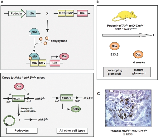
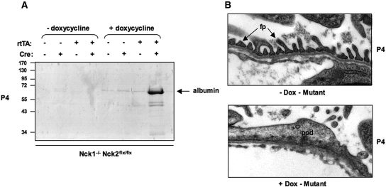
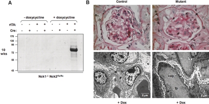
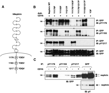
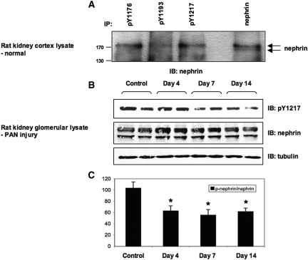
Within the glomerulus, the scaffolding protein nephrin bridges the actin-rich foot processes that extend from adjacent podocytes to form the slit diaphragm. Mutations affecting a number of slit diaphragm proteins, including nephrin, cause glomerular disease through rearrangement of the actin cytoskeleton and disruption of the filtration barrier. We recently established that the Nck family of Src homology 2 (SH2)/SH3 cytoskeletal adaptor proteins can mediate nephrin-dependent actin reorganization. Formation of foot processes requires expression of Nck in developing podocytes, but it is unknown whether Nck maintains podocyte structure and function throughout life. Here, we used an inducible transgenic strategy to delete Nck expression in adult mouse podocytes and found that loss of Nck expression rapidly led to proteinuria, glomerulosclerosis, and altered morphology of foot processes. We also found that podocyte injury reduced phosphorylation of nephrin in adult kidneys. These data suggest that Nck is required to maintain adult podocytes and that phosphotyrosine-based interactions with nephrin may occur in foot processes of resting, mature podocytes.


Figures

Figure 1.
Figure 1.
Generation of a conditional knockout of Nck in adult podocytes, using the Tet-On system. (A) Binary transgenic system in which one transgene places the rtTA under the control of the podocyte-specific podocin promoter, while a second transgene uses tetO promoter elements upstream of a minimal CMV promoter to drive expression of Cre recombinase. rtTA requires the tetracycline analog doxycycline to bind tetO and produce Cre protein. These transgenes are introduced into mice homozygous for the Nck1-null (-) allele and the Nck2 floxed (flx) allele. Upon doxycycline treatment, Cre recombinase induces recombination between the loxP sites on the Nck2 allele, resulting in deletion of Nck2 selectively in podocytes of Nck1 null mice. Since Cre is not produced in any other cell types, they will continue to express Nck2 protein. Loss of Nck2 in podocytes is irreversible. (B) Mutant mice were exposed to doxycycline at E13.5 to delete Nck2 in developing podocytes or at 4 wk of age to delete Nck2 in established podocytes. (C) Binary transgenic mice were intercrossed with the Z/EG reporter strain to demonstrate podocyte-restricted expression of Cre recombinase. Cre-mediated excision results in expression of GFP, which is visualized by staining kidney sections with anti-GFP antibodies.
Figure 2.
Figure 2.
Doxycycline-mediated Cre induction and loss of Nck expression in developing podocytes results in nephrotic syndrome. (A) SDS-PAGE analysis of urine from littermates at postnatal day 4 (P4). All mice are Nck1-/- Nck2flx/flx, and carrying neither, one or both Podocin-rtTA and tetO-Cre transgenes, as indicated. Double transgenic Nck1-/- Nck2flx/flx mice (referred to as “mutant”) develop albuminuria when exposed to doxycycline in utero at E13.5. Proteinuria is not seen in untreated mutants, or single transgenic control littermates exposed to doxycycline. (B) Transmission electron micrographs of the glomerular filtration barrier in mutant mice exposed to doxycycline in utero or left untreated. Well-separated foot processes (fp) with intervening slit diaphragms are observed in untreated mutant mice (upper panel), whereas mice exposed to doxycycline to induce Cre-mediated Nck2 excision show flattened podocytes (pod) with an absence of slit diaphragms (lower panel).
Figure 3.
Figure 3.
Genetic deletion of Nck expression in established podocytes demonstrates an ongoing requisite role for Nck signaling at the slit diaphragm. (A) SDS-PAGE shows proteinuria in mutant mice exposed to doxycycline at 4 wk of age to induce Nck2 deletion, but not in untreated mutants, or single transgenic control littermates. All mice are Nck1-/- Nck2flx/flx, and carrying neither, one or both Podocin-rtTA and tetO-Cre transgenes, as indicated. Mice were analyzed at 10 wk of age. (B) PAS staining of kidney sections reveals prominent glomerulosclerosis in mutant mice, with tuft adhesions to Bowman's capsule (arrow) and protein deposits in the tubules (asterisk). Electron micrographs show widespread podocyte (pod) effacement with electron dense areas along the basement membrane consistent with collapsed actin (actin), although distinct foot processes (fp) can still be observed around some capillary (cap) loops. Normal glomerular morphology and foot process architecture in a doxycycline-treated littermate control is shown for comparison.
Figure 4.
Figure 4.
Early alterations in glomerular morphology and function upon loss of Nck expression in adult podocytes. Adult mice (4 wk of age) were exposed to doxycycline and analyzed after 1 or 2 wk. (A) An increase in proteinuria is seen at these timepoints in mutant mice, but not in doxycycline-treated littermate controls. (B) Foot process architecture is maintained in control mice exposed to doxycycline. (C and D) By contrast, conditional Nck mutants exhibit focal areas of foot process fusion and broadening (arrow) at 1 wk after doxycycline exposure, and these ultrastructural changes are widespread after 2 wk of induction with areas of diffuse foot process fusion. Fenestrated endothelial cells are relatively spared in mutant mice.
Figure 5.
Figure 5.
Conserved tyrosine residues on nephrin are recognized by phospho-specific antibodies. (A) The intracellular region of human nephrin contains a series of conserved tyrosine residues within YDxV motifs that can bind the Nck SH2 domain. (B) Peptides encompassing each tyrosine phosphorylated YDxV motif on nephrin were used to immunize rabbits and generate rabbit monoclonal antibodies (RAbMAbs). HEK 293T cells were transfected with a series of GFP-tagged chimeric human nephrin proteins with tyrosine (Y)-to-phenylalanine (F) substitutions at amino acid positions 1176, 1193, and 1217. Nephrin phosphorylation was induced by CD16 clustering, and lysates were immunoprecipitated (IP) with GFP antibodies to isolate nephrin. Separated proteins were immunoblotted (IB) with affinity-purified phospho-nephrin antibodies or GFP. Both the nephrin pY1193 and pY1217 antibodies appear to be specific to each respective Tyr residue, while the pY1176 antibody appears to recognize YDEV (1176 and also 1193 to a limited extent) but not YDQV (1217) sites. None of the antibodies could detect the Y3F triple mutant, indicating their specificity for phosphorylated YDxV sites on nephrin. Exposure time for the pY1176 and pY1193 blots is 30 min, and exposure time for the pY1217 blot is 10 min. (C) HEK 293T cells expressing wildtype (WT) chimeric nephrin were stimulated with anti-CD16 or left unstimulated, and lysates were immunoprecipitated (IP) with each phospho-nephrin antibody or GFP. Separated proteins were immunoblotted (IB) with GFP or phosphotyrosine (pY) antibodies. Nephrin clustering results in increased tyrosine phosphorylation on each YDxV site.
Figure 6.
Figure 6.
Nephrin is phosphorylated on YDxV sites in normal adult kidneys and a decrease is associated with podocyte injury. (A) Adult rat kidney cortex lysates were immunoprecipitated (IP) with each phospho-nephrin antibody or pan-nephrin, and separated proteins were immunoblotted (IB) with nephrin antibodies. Endogenous nephrin appears as a doublet of 180 and 170 kD, and each of the phospho-nephrin antibodies detected the 180-kD tyrosine-phosphorylated upper species. (B) Glomerular lysates from rats with PAN nephrosis at various time-points or untreated control rats were immunoblotted with phospho-nephrin or nephrin antibodies. We utilized the pY1217 antibody as it displays superior sensitivity. Equivalent amounts of lysate are indicated by tubulin. Data shown are representative of four independent experiments. (C) Densitometric quantitation of phosphorylated nephrin normalized to total nephrin (p-nephrin/nephrin) indicates a decrease in nephrin phosphorylation on the YDQV site upon podocyte injury. *P < 0.05 versus control, n = 4.

Comment in

References

    1. Pavenstadt H, Kriz W, Kretzler M: Cell biology of the glomerular podocyte. Physiol Rev 83: 253–307, 2003 - PubMed
    1. Michaud JL, Kennedy CR: The podocyte in health and disease: Insights from the mouse. Clin Sci (Lond) 112: 325–335, 2007 - PubMed
    1. Wiggins RC: The spectrum of podocytopathies: A unifying view of glomerular diseases. Kidney Int 71: 1205–1214, 2007 - PubMed
    1. Patrakka J, Tryggvason K: Nephrin—A unique structural and signalling protein of the kidney filter. Trends Mol Med 13: 396–403, 2007 - PubMed
    1. Lahdenpera J, Kilpelainen P, Liu XL, Pikkarainen T, Reponen P, Ruotsalainen V, Tryggvason K: Clustering-induced tyrosine phosphorylation of nephrin by Src family kinases. Kidney Int 64: 404–413, 2003 - PubMed

Publication types

MeSH terms