. 2009 Aug;20(8):1714-23.
doi: 10.1681/ASN.2008060640. Epub 2009 May 14.

The CXCL12 (SDF-1)/CXCR4 axis is essential for the development of renal vasculature

Affiliations

The CXCL12 (SDF-1)/CXCR4 axis is essential for the development of renal vasculature

Yoshitsugu Takabatake et al. J Am Soc Nephrol. 2009 Aug.

Abstract

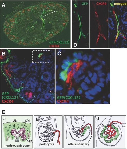
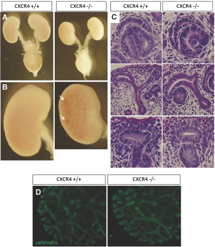
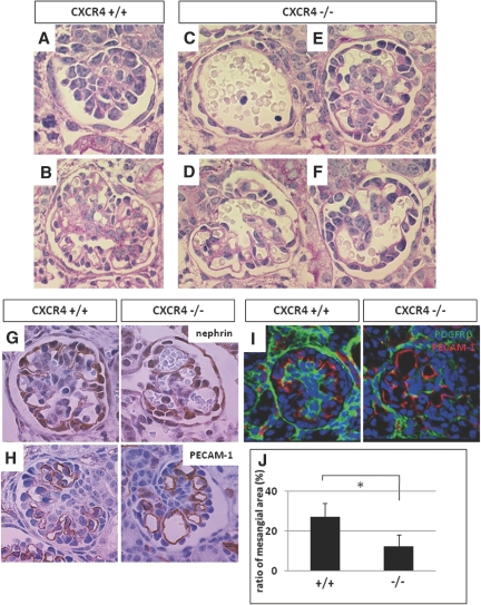
CXC chemokine ligand 12 (CXCL12; stromal cell-derived factor 1) is a unique homeostatic chemokine that signals through its cognate receptor, CXCR4. CXCL12/CXCR4 signaling is essential for the formation of blood vessels in the gastrointestinal tract during development, but its contribution to renal development remains unclear. Here, we found that CXCL12-secreting stromal cells surround CXCR4-positive epithelial components of early nephrons and blood vessels in the embryonic kidney. In glomeruli, we observed CXCL12-secreting podocytes in close proximity to CXCR4-positive endothelial cells. Both CXCL12- and CXCR4-deficient kidneys exhibited identical phenotypes; there were no apparent abnormalities in early nephrogenesis or in differentiation of podocytes and tubules, but there was defective formation of blood vessels, including ballooning of the developing glomerular tuft and disorganized patterning of the renal vasculature. To clarify the relative importance of different cellular defects resulting from ablation of CXCL12 and CXCR4, we established endothelial cell-specific CXCR4-deficient mice, which recapitulated the renal phenotypes of conventional CXCR4-deficient mice. We conclude that CXCL12 secreted from stromal cells or podocytes acts on endothelial cells to regulate vascular development in the kidney. These findings suggest new potential therapeutic targets for remodeling the injured kidney.


Figures

Figure 1.
Figure 1.
Expression of CXCL12 in the developing kidney using CXCL12/GFP knock-in mice at E15.5. (A) Appearance of a whole section of the kidney at low magnification. (B) The section was immunostained red (Alexa555) for calbindin, a UB marker. GFP was expressed in the stromal cells surrounding developing nephrons (UB, CM [arrows], and pretubular aggregates [PA; arrowheads]). (C and D) Another section was immunostained with Alexa555-labeled anti-VEGFR2 antibody, a marker for endothelial cells. (C) CXCL12 was expressed in some developing podocytes (arrows) in the primitive glomeruli. (D) All podocytes express CXCL12 in the mature glomeruli. Magnifications: ×100 in A; ×400 in B; ×600 in C and D.
Figure 2.
Figure 2.
Expression of CXCR4 in the developing kidney. CXCR4 was stained red (Alexa555) in the kidney sections of wild-type mice at E13.5 (A) and E17.5 (B through G). (A and B) Whole sections of the kidneys under low magnification revealed that CXCR4 is mainly expressed in the nephrogenic zone. (C) Co-immunostaining for cited1, a marker for CM, in green (Alexa488; left: CXCR4, middle: merged image) shows that CXCR4 is expressed in CM (arrows in inset) and PA (arrowheads in inset). Sections of CXCR4 null kidney are shown as negative control for CXCR4 staining (right). (D through F) Another section was co-immunostained for VEGFR2 in green (Alexa488; left: CXCR4, right: merged image). (D) CXCR4 was first detected only weakly in the endothelial cells in the cleft of the comma-shaped body (arrows) and more intensely in those at a basal position (arrowheads). (E) CXCR4 was detected in arterioles leading to the primitive glomeruli (arrows). Weak expression was detected in a stalk of glomerular endothelial cells (arrowheads). (F) In more mature glomeruli, CXCR4 was expressed in many glomerular endothelial cells. (G) Co-immunostaining for renin in green (Alexa488) shows that renin is expressed in pericytes surrounding CXCR4-positive endothelial cells, suggesting that CXCR4-positive vessels are afferent arterioles. Glm, glomerulus. Magnifications: ×100 in A and B; ×400 in C and E; ×600 in D, F, and G.
Figure 3.
Figure 3.
Spatial relationship between CXCL12 and CXCR4 in developing kidney. Kidney sections from CXCL12/GFP knock-in mice were stained red (Alexa555) for CXCR4. (A and B) Appearance of whole kidney section (A) and magnified image (B) demonstrates that CXCL12-positive stromal cells surround CXCR4-positive developing nephrons. (C) Higher magnification of the glomerulus in the dotted lines in B suggests that some podocytes express CXCL12 and that glomerular endothelial cells just adjacent to the podocytes express CXCR4. (D) Higher magnification of the region in the dotted lines in A of GFP (left) and CXCR4 staining (middle) in the interlobular arteries. Merged image (right) indicates that at least some interlobular arteries express both CXCL12 and CXCR4. (E) Summary of the expression of CXCL12 (green) and CXCR4 (red) in the developing early nephron (a) and glomeruli (b through d). See the Discussion section for more details. Magnifications: ×100 in A; ×400 in B; ×600 in C and D.
Figure 4.
Figure 4.
Analysis of CXCR4 null kidneys. (A and B) Stereoscopic appearances of wild-type (left) and CXCR4 null (right) urinary organs. Kidneys, urinary tracts, and bladders from mice at E17.5 are shown after removal of genital organs. (A) CXCR4 null kidneys are smaller, but the urinary tract and bladder show no macroscopic abnormalities. (B) Magnified images of CXCR4 null kidneys show petechial hemorrhages (arrows). (C) Assessment of early development of nephrons. Periodic acid-Schiff (PAS)-stained sections of wild-type (left) or CXCR4 null (right) kidneys at E13.5 are shown. Early nephrogenesis, including formation of comma- or S-shaped bodies (top), UB branching and formation of renal vesicles (middle), and aggregations of metanephric mesenchyme (lower), was not affected by ablation of CXCR4. (D) Branching morphogenesis of CXCR4 null kidneys. Kidneys were resected from wild-type or CXCR4 KO mice at E12.5 and cultured on Transwell for 48 h. They were then whole mount–immunostained for calbindin, revealing that UB branching was not affected by the ablation of CXCR4. Magnifications: ×10 in A and B; ×200 in C and D.
Figure 5.
Figure 5.
Analysis of glomerulogenesis in CXCR4 null kidneys. (A through F) Deficiencies in glomerulogenesis in CXCR4 null kidneys. PAS-stained images of premature to mature glomeruli of wild-type (A and B) or CXCR4 KO (C through F) mice at E17.5 are shown. Compared with wild-type kidney, CXCR4 null kidneys display abnormal glomerulogenesis: Some glomeruli consist of a single capillary tuft surrounded by a single array of cells (C), and other glomeruli show ballooning of capillary tufts to various extents (D through F). (G through I) Expression of markers for cell lineages in wild-type (left) and CXCR4 null (right) glomeruli at E17.5. Glomeruli were immunostained for nephrin for podocytes (G), PECAM-1 for endothelial cells (H), and PDGFRβ for mesangial cells (I). (J) Analysis of the ratio of the PDGFRβ-positive area (mesangial cells area) to the total glomerular area in wild-type and CXCR4 null kidneys. The ratio is expressed as the mean ± SD. *P < 0.05 versus wild-type glomeruli. Magnification, ×1000.
Figure 6.
Figure 6.
Electron microscopic analysis of CXCR4 null glomeruli. Low-magnification (top) and high-magnification (middle) images show foot process effacement (arrows) after CXCR4 ablation, although endothelial cells and podocytes are securely attached to the GBM. (Bottom) There are fewer fenestrations in CXCR4 null glomeruli than in normal glomeruli (arrows). Magnifications: ×2000 in top; ×10,000 in middle and bottom.
Figure 7.
Figure 7.
Assessment of vascular patterning of whole kidneys from CXCR4 KO mice. (A through C) Whole-mount immunostaining for PECAM-1 was performed using kidneys from wild-type (left) and CXCR4 KO (right) mice at E13.5 (A), E15.5 (B), and E17.5 (C). Positive staining was visualized by the horseradish peroxidase–DAB method. Magnified images are also presented in the insets. In the CXCR4 null kidney, regular vascular pattering is disrupted, with narrowing (arrows) or dilation (arrowheads) observed at all stages. Magnification, ×10.
Figure 8.
Figure 8.
Analysis of the kidneys from endothelial cell–specific CXCR4 KO mice. (A through F) PAS-stained kidney sections of CXCR4 flox/+ (as a control; A) and Tie2-Cre CXCR4 flox/flox mice (B through F) 3 d after birth. (A and B) Low-magnification images. Arrows indicate abnormally dilated tubules. (C and D) Magnified view of B (right). Proximal tubules are dilated and flattened in shape with vacuolations (arrows). (E and F) Glomeruli of Tie2-Cre CXCR4 flox/flox mice exhibit ballooning of the capillary tuft, identical to conventional CXCR4 null glomeruli. Some endothelial cells are detached from the GBM (arrows). (G) Electron microscopic images of glomeruli from Tie2-Cre CXCR4 flox/flox mice. Endothelial cells detached from the GBM are shown magnified (middle and right). Magnifications: ×100 in A and B; ×200 in C; ×1000 in D through F; ×2000 in G, left; ×10,000 in G, middle and right.

Comment in

References

    1. Vaughan MR, Quaggin SE: How do mesangial and endothelial cells form the glomerular tuft? J Am Soc Nephrol 19: 24–33, 2008 - PubMed
    1. Nagasawa T: A chemokine, SDF-1/PBSF, and its receptor, CXC chemokine receptor 4, as mediators of hematopoiesis. Int J Hematol 72: 408–411, 2000 - PubMed
    1. Ratajczak MZ, Zuba-Surma E, Kucia M, Reca R, Wojakowski W, Ratajczak J: The pleiotropic effects of the SDF-1-CXCR4 axis in organogenesis, regeneration and tumorigenesis. Leukemia 20: 1915–1924, 2006 - PubMed
    1. Tachibana K, Hirota S, Iizasa H, Yoshida H, Kawabata K, Kataoka Y, Kitamura Y, Matsushima K, Yoshida N, Nishikawa S, Kishimoto T, Nagasawa T: The chemokine receptor CXCR4 is essential for vascularization of the gastrointestinal tract. Nature 393: 591–594, 1998 - PubMed
    1. Nagasawa T, Kikutani H, Kishimoto T: Molecular cloning and structure of a pre-B-cell growth-stimulating factor. Proc Natl Acad Sci U S A 91: 2305–2309, 1994 - PMC - PubMed

Publication types

LinkOut - more resources