. 2009 Jun 2;106(22):8941-6.
doi: 10.1073/pnas.0812469106. Epub 2009 May 14.

LKB1 regulates polarity remodeling and adherens junction formation in the Drosophila eye

Affiliations

LKB1 regulates polarity remodeling and adherens junction formation in the Drosophila eye

Nancy Amin et al. Proc Natl Acad Sci U S A. .

Abstract

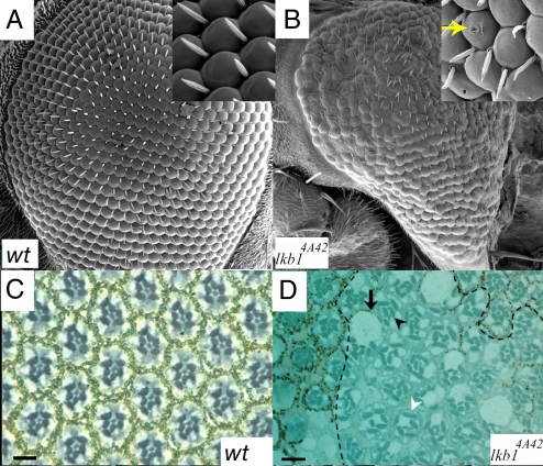
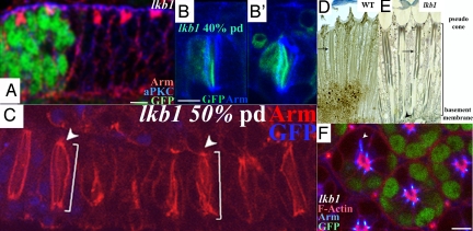
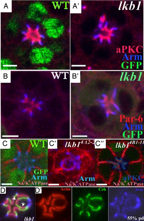
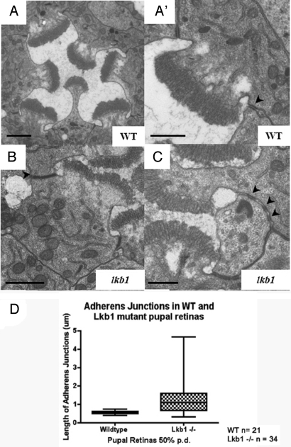
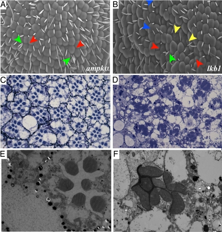
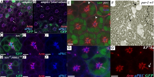
The serine-threonine kinase LKB1 regulates cell polarity from Caenorhabditis elegans to man. Loss of lkb1 leads to a cancer predisposition, known as Peutz-Jeghers Syndrome. Biochemical analysis indicates that LKB1 can phosphorylate and activate a family of AMPK- like kinases, however, the precise contribution of these kinases to the establishment and maintenance of cell polarity is still unclear. Recent studies propose that LKB1 acts primarily through the AMP kinase to establish and/or maintain cell polarity. To determine whether this simple model of how LKB1 regulates cell polarity has relevance to complex tissues, we examined lkb1 mutants in the Drosophila eye. We show that adherens junctions expand and apical, junctional, and basolateral domains mix in lkb1 mutants. Surprisingly, we find LKB1 does not act primarily through AMPK to regulate cell polarity in the retina. Unlike lkb1 mutants, ampk retinas do not show elongated rhabdomeres or expansion of apical and junctional markers into the basolateral domain. In addition, nutrient deprivation does not reveal a more dramatic polarity phenotype in lkb1 photoreceptors. These data suggest that AMPK is not the primary target of LKB1 during eye development. Instead, we find that a number of other AMPK-like kinase, such as SIK, NUAK, Par-1, KP78a, and KP78b show phenotypes similar to weak lkb1 loss of function in the eye. These data suggest that in complex tissues, LKB1 acts on an array of targets to regulate cell polarity.


Conflict of interest statement

The authors declare no conflict of interest.

Figures

Fig. 1.
Fig. 1.
Mutation of lkb1 disrupts eye development. (A) SEMs of a wild-type eye show a regular array of ommatidia and bristles. (B) SEMs of the lkb14A4-2 eye; defects include fused ommatidia, missing and excess bristles, and disorganized bristles. The eye is also smaller, rougher, and misshapen with “pitting” of the surface (Inset). (C) Light micrograph of a 1-μm cross section through a wild-type retina reveals a stereotypical arrangement of photoreceptors and ommatidia. (D) lkb1 clones are identified by the lack of pigment and are contained within dashed lines. Loss of lkb1 leads to a loss of photoreceptors (black arrowhead), misshapen rhabdomeres (white arrowhead), and enlarged cell bodies (black arrow). (Scale bars, 10 μm.)
Fig. 2.
Fig. 2.
lkb1 affects polarity at pupal stages and PRCs extend properly. (A, B, and F) lkb14B1-11. (C and D) lkb14A42. (A) Epithelial polarity is maintained in lkb1 third-instar clones; aPKC (blue) and Arm (red) are correctly localized in lkb1 tissue (marked by loss of GFP). (B) Junctional membranes in 40% pd lkb1 PRCs do not fragment, as shown by continuous Arm (blue) staining. (C) At 50% pd, PRCs undergo normal proximodistal extension (white scale bar), and rhabdomere feet remain attached to the basement membrane (white arrowhead). (D and E) Adult longtitudinal sections; lkb1 rhabdomeres (E) show breaks throughout the proximal–distal length of the rhabdomere, but PRCs extend normally to the basement membrane (black arrowhead). lkb1 rhabdomeres show “waviness” of the lateral membranes (arrow in E) compared with wild type (arrow in D). (F) Confocal section although a 40% pd, lkb1 mosaic eye, showing a wild-type ommatidia (green) alongside a mutant ommatida. Arm staining (blue) extends in cells lacking lkb1. (Scale bars, 5 μm.)
Fig. 3.
Fig. 3.
lkb1 loss of results in polarity defects in the pupal retina. GFP (green) marks wild-type tissue in A and C and mutant tissue in B. lkb14B1-11 (A–C) and lkb14A42 (D and E). Apical markers aPKC and Par-6 (red) show expansion into the basolateral domain in lkb1 mutant clones. (B′) The junctional marker Arm (blue) also shows aberrant expansion into the basolateral domain. (C′) The basolateral marker Na+/K+ ATPase (red) also mislocalizes to the apical membrane in lkb14B1-11 mosaic PRCs. (C″) lkb14A42 mosaic retinas show a more severe phenotype, where Na+/K+ ATPase (red) can be found in a ring like structures overlapping Arm (blue). (D′) Extra membrane domains are sometimes observed, e.g., 3 subapical domains, 2 apical domains (white arrowheads). (Scale bars, 5 μm.)
Fig. 4.
Fig. 4.
Adherens junctions are longer, more numerous, and mislocalized in lkb1 photoreceptor cells. (A and A′) Ultrathin sections (70 nm) of a wild-type ommatidium at 50% pd. AJ in wild-type PRCs occupy an apicolateral position in the cell, and each cell has 2 AJs (black arrowhead) of uniform length (0.5 μm). (B–C) lkb1 AJs are frequently longer (white arrowhead in B) and sometimes disjointed (black arrowheads in C). (Scale bars, 1 μm in A and B and 0.5 μm in A′ and C.) (D) Box-plot analysis of lkb1 AJ length in PRCs in 50% pd pupal retinas. The average length of AJs in lkb1 PRC increased to 1.28 μm. AJs also exhibit an increased range in length. Smaller junction lengths may indicate fragmented AJs.
Fig. 5.
Fig. 5.
lkb1 and ampkα mutant adult eyes. (A–F) SEMs of adult eyes. ampkα and lkb1 mutant eyes are “rough” (A and B). lkb1 mutant ommatidia show pitting of the surface (yellow arrowhead in B) and rhabdomere fusion (blue arrowhead in B) that is not observed in ampkα mutant eyes (A). Bristles missing between ommatidia (red arrows in A and B) or duplicated (green arrows in A and B) are frequently observed. (C and D) Light micrographs of ampkα (C) and lkb1 (D) mutant eyes. Both ampkα and lkb1 mutant eyes show enlarged cell bodies (C and D), whereas only lkb1 shows elongation of rhabdomeres (D compared with C). (E–F) TEM of ampkα (E) and lkb1 (F) mutants. R7 is sometimes absent in sections of ampkα mutants (E), whereas the rhabdomere membrane is often enlarged in lkb1 mutants (F).
Fig. 6.
Fig. 6.
ampk−/− does not alter Arm localization, whereas KP78a loss phenocopies lkb1. GFP (green) marks the wild-type tissue in A, B, and E, MARCM clones in C and D, and flp-out clones in G and H. ampkα3 mutant clones do not phenocopy lkb1 clones raised on normal food (A) or starvation food (B). ampkα3 mutant PRCs show discrete Arm (red) localization (arrows in A and B). Expression of MRLCEE in lkb1 clones does not rescue lkb1 polarity defects (D). (C) MARCM lkb1X5 clones show basolateral spreading of aPKC (blue) and Arm (red). (D) MARCM lkb1X5 clones expressing MRLCEE do not show a rescue of the basolateral spreading of apical (aPKC, blue) and junctional markers (Arm, red). (E) par1w3 clones show expansion of Arm (red) toward the basolateral domain of PRCs (arrowheads). (F) par1w3 clones (lack of pigment cells) show elongated rhadomeres (arrowhead) and enlarged cells bodies (arrow); phenotypes also characteristic of lkb1 loss of function in the eye. (G) Expression of KP78a RNAi results in basolateral spreading of apical (aPKC, blue) and junctional markers (Arm, red). (Scale bars, 5 μm.)

References

    1. Lizcano JM, et al. LKB1 is a master kinase that activates 13 kinases of the AMPK subfamily, including MARK/PAR-1. EMBO J. 2004;23:833–843. - PMC - PubMed
    1. Marignani PA. LKB1, the multitasking tumour suppressor kinase. J Clin Pathol. 2005;58:15–19. - PMC - PubMed
    1. Watts JL, Morton DG, Bestman J, Kemphues KJ. The C. elegans par-4 gene encodes a putative serine–threonine kinase required for establishing embryonic asymmetry. Development. 2000;127:1467–1475. - PubMed
    1. Morton DG, Roos JM, Kemphues KJ. par-4, a gene required for cytoplasmic localization and determination of specific cell types in Caenorhabditis elegans embryogenesis. Genetics. 1992;130:771–790. - PMC - PubMed
    1. Martin SG, St Johnston D. A role for Drosophila LKB1 in anterior–posterior axis formation and epithelial polarity. Nature. 2003;421:379–384. - PubMed

Publication types

MeSH terms

Substances

LinkOut - more resources