. 2009 May 26;106(21):8766-71.
doi: 10.1073/pnas.0903499106. Epub 2009 May 14.

Neurotensin induces IL-6 secretion in mouse preadipocytes and adipose tissues during 2,4,6,-trinitrobenzensulphonic acid-induced colitis

Affiliations

Neurotensin induces IL-6 secretion in mouse preadipocytes and adipose tissues during 2,4,6,-trinitrobenzensulphonic acid-induced colitis

Hon-Wai Koon et al. Proc Natl Acad Sci U S A. .

Abstract

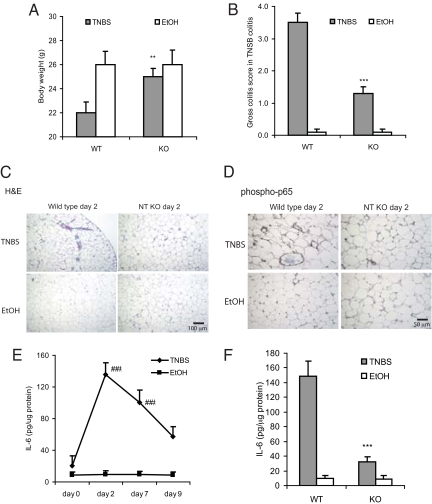
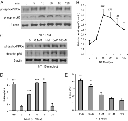
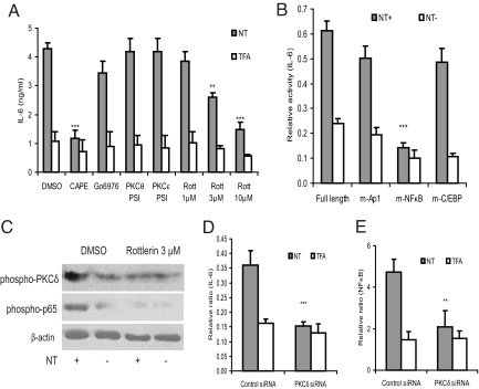
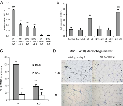
Mesenteric fat is known to undergo inflammatory changes after 2,4,6,-trinitrobenzensulphonic acid (TNBS)-induced colitis. Neurotensin (NT) and neurotensin receptor 1 (NTR1) have been shown to play a major role in the pathogenesis of intestinal inflammation. This led us to explore whether NT and NTR1 are expressed in the mesenteric fat depots during TNBS-induced colitis and whether NT participates in the increased interleukin (IL)-6 secretion in this inflammatory response. TNBS-induced inflammation in the colon increases NT and NTR1 expression in mesenteric adipose tissues, including mesenteric preadipocytes. Compared with wild-type mice, NT knockout (KO) mice have reduced TNBS-induced colitis accompanied by diminished inflammatory responses in mesenteric adipose tissue. Specifically, IL-6 and p65 phosphorylation levels in mesenteric fat of NT KO mice are also reduced compared with wild-type mice. Mouse 3T3-L1 preadipocytes express NTR1 and its expression is increased after stimulation of preadipocytes with proinflammatory cytokines. NT stimulation of 3T3-L1 preadipocytes overexpressing NTR1 causes PKCdelta phosphorylation and IL-6 secretion in a time- and dose-dependent fashion. Moreover, NT-mediated IL-6 expression is nuclear factor-kappaB and PKCdelta dependent. We also found that supernatants from NT-exposed 3T3-L1-NTR1 preadipocytes and mesenteric fat obtained from wild-type mice 2 days after TNBS administration stimulate an IL-6-dependent macrophage migration measured by a macrophage migration assay, whereas this response is reduced when mesenteric fat from NT KO mice is used. These results demonstrate an important role for NT in acute colitis and adipose tissue inflammation associated with experimental colitis that involves direct NT proinflammatory responses in preadipocytes.


Conflict of interest statement

The authors declare no conflict of interest.

Figures

Fig. 1.
Fig. 1.
Increased expression of NT and NTR1 in mesenteric fat of mice during TNBS-induced colitis: Eight-week-old male C57BL/6 mice (six mice per group) were treated with either TNBS or 30% ethanol (vehicle) for 0, 2, 7, and 9 days. Protein levels of NTR1 in mesenteric fat were measured by Western blot (A). (Right lane) Migration of prestained molecular weight protein standards (k = 1000 daltons) as described in Materials and Methods. (B) Densitometric measurements of NTR1 Western blots were normalized by β-actin. (C) Peptide levels of NT in mesenteric fat were measured by enzyme-linked immunosorbent assay (ELISA) (***P < 0.001 vs. day 0). Immunofluorescence staining for NTR1 (D), NT (E) as well as Pref-1 (a preadipocyte marker), and nuclear stain DAPI (D, E) of mesenteric fat from mice with TNBS colitis (day 2) and ethanol-treated control mice. Overlapping expression of NTR1/NT (Texas Red) with Pref-1 (FITC green) indicates preadipocytes expressing NTR1 or NT (yellow); magnification ×200.
Fig. 2.
Fig. 2.
NT KO mice are protected from colonic tissue damage, adipose inflammation, and NF-κB activation caused by TNBS-induced colitis. Homozygous NT KO and wild-type mice were treated with TNBS for 2 days and body weight changes (A) and gross colitis score (B) were measured. ***P < 0.001, **P < 0.01 vs. wild-type, TNBS-treated mice. Mesenteric fat tissues from the same mouse groups were either stained with hematoxylin and eosin (C) or an antibody against phosphorylated p65 (D). (E) Mesenteric fat IL-6 levels of wild-type mice throughout the course of TNBS treatment were measured. ###P < 0.001 vs. day 0 of TNBS treated mice. (F) Mesenteric fat IL-6 levels on day 2 post-TNBS from wild-type and NT KO mice were measured. All experiments are representative of six mice per group. *** P < 0.001 vs. wild-type mice with TNBS treatment. Magnification ×100 (C) or ×200 (D).
Fig. 3.
Fig. 3.
NT induces IL-6 secretion in 3T3-L1-NTR1 preadipocytes: (A, C) Cultured 3T3-L1-NTR1 3T3-L1 preadipocytes were exposed to different concentrations of NT at various time intervals. Cells were lysed and equal amounts of proteins were blotted for detection of phosphorylated PKCδ and p65, or β-actin. (B) Densitometric analyses of phospho-p65 signal normalized to β-actin signal. ###P < 0.001, ##P < 0.01 vs. 0 minute. Cultured 3T3-L1-NTR1 preadipocytes were exposed to NT (10 nM) or vehicle trifluoroacetic acid 0.1% (TFA) for 0–24 hours (D), and at various doses (0–100 nM) for 8 hours (E). Treatment with 1 μM proinflammatory phorbol ester phorbol-12-myristate-13-acetate (PMA) for 8 hours served as a positive control. Conditioned media were collected for mouse IL-6 ELISA. +++P < 0.001 vs. 0 h. ***P < 0.001, **P < 0.01 vs. TFA control group. Results are representative of three independent experiments.
Fig. 4.
Fig. 4.
NT-induced IL-6 secretion in 3T3-L1-NTR1 mouse preadipocytes involves PKCδ- and NF-κB–dependent pathways: (A) 3T3-L1-NTR1 preadipocytes were pretreated with dimethyl sulfoxide (DMSO; vehicle), CAPE (10 μM), Ca2+ dependent PKC inhibitor Go6976 (10 μM), Rottlerin (1–10 μM), PKCθ pseudosubstrate inhibitor (PKCθ PSI; 10 μM) or PKCε pseudosubstrate inhibitor (PKCε PSI; 10 μM) for 30 minutes, followed by NT (10 nM) or TFA (vehicle) for 8 hours. Conditioned media were then collected for mouse IL-6 ELISA. Results were representative of three independent experiments. (B) 3T3-L1-NTR1 preadipocytes were transfected with either full-length IL-6 promoter construct or IL-6 promoter constructs containing mutations in the NF-κB (m-NF-κB), AP1 (m3/m5-Ap1), or C/EBP (mC/EBP) transcription binding sites, followed by NT (10 nM) treatment for 8 hours. Cell lysates were then used to perform luciferase reporter assays. ***P < 0.001 vs. full-length group. (C) 3T3-L1-NTR1 preadipocytes were pretreated with DMSO or Rottlerin (3 μM) for 30 minutes, followed by NT exposure (10 nM) for 30 minutes. Cells were lysed, and equal amounts of protein were used to detect phospho-PKCδ, phospho-p65, and β-actin. (D, E) 3T3-L1-NTR1 preadipocytes were co-transfected with control siRNA or PKCδ siRNA together with a IL-6 promoter (D) and NF-κB luciferase construct (E), followed by exposure to NT (10 nM) for 8 hours. Cell lysates were used to perform IL-6/NF-κB luciferase reporter assay. ***P < 0.001 vs. control siRNA transfected NT-treated group. Results are representative of three independent experiments.
Fig. 5.
Fig. 5.
NT stimulates preadipocyte-dependent macrophage migration via IL-6 secretion. (A) Mesenteric fat extracts from wild-type and NT KO mice were was placed in the lower compartment of modified Boyden chambers together with either anti-mouse IL-6–neutralizing IgG or control IgG. Murine macrophage Raw264.7 cells were then seeded into the upper compartment; after 8 hours, cells that had migrated to the membrane were stained and lysed, and cell migration was determined by absorbance measurements at 560 nm. ###P < 0.001 vs. Wt+EtOH+IgG; ***P < 0.001 vs. Wt+TNBS+IgG. (B) 3T3-L1-NTR1 preadipocytes, placed onto the lower compartment of modified Boyden chambers, were treated with IgG, anti-IL-6 antibody or NTR1 receptor antagonist SR48692 (2 μM) followed by exposure to NT (10 nM) or TFA (1 μl) for 8 hours and measurement of macrophage cell migration as described in (A). ***P < 0.01 vs. Con+IgG; †††P < 0.01 vs. NT+IgG; ###P < 0.001 vs. Con+IgG. Results are representative of six independent experiments. (C) Homozygous NT KO and wild-type mice were treated with TNBS for 2 days. RNA was isolated from mesenteric fat and macrophage-specific EMR1 (F4/80) mRNA was quantitated by real-time RT-PCR. (D) Mesenteric fat tissues from the same mouse groups were also stained with an antibody against EMR1. All experiments are representative of six mice per group. ***P < 0.01 vs. wild-type mice. Magnification ×200.

References

    1. Kershaw EE, Flier JS. Adipose tissue as an endocrine organ. J Clin Endocrinol Metab. 2004;89:2548–2556. - PubMed
    1. Xu H, et al. Chronic inflammation in fat plays a crucial role in the development of obesity-related insulin resistance. J Clin Invest. 2003;112:1821–1830. - PMC - PubMed
    1. Weisberg SP, et al. Obesity is associated with macrophage accumulation in adipose tissue. J Clin Invest. 2003;112:1796–1808. - PMC - PubMed
    1. Desreumaux P, et al. Inflammatory alterations in mesenteric adipose tissue in Crohn's disease. Gastroenterology. 1999;117:73–81. - PubMed
    1. Schaffler A, Scholmerich J, Buchler C. Mechanisms of disease: Adipocytokines and visceral adipose tissue—emerging role in nonalcoholic fatty liver disease. Nat Clin Pract Gastroenterol Hepatol. 2005;2:273–280. - PubMed

Publication types

MeSH terms

LinkOut - more resources