. 2009 Jun 5;104(11):1302-12.
doi: 10.1161/CIRCRESAHA.109.197830. Epub 2009 May 14.

Activated forms of VEGF-C and VEGF-D provide improved vascular function in skeletal muscle

Affiliations

Activated forms of VEGF-C and VEGF-D provide improved vascular function in skeletal muscle

Andrey Anisimov et al. Circ Res. .

Abstract

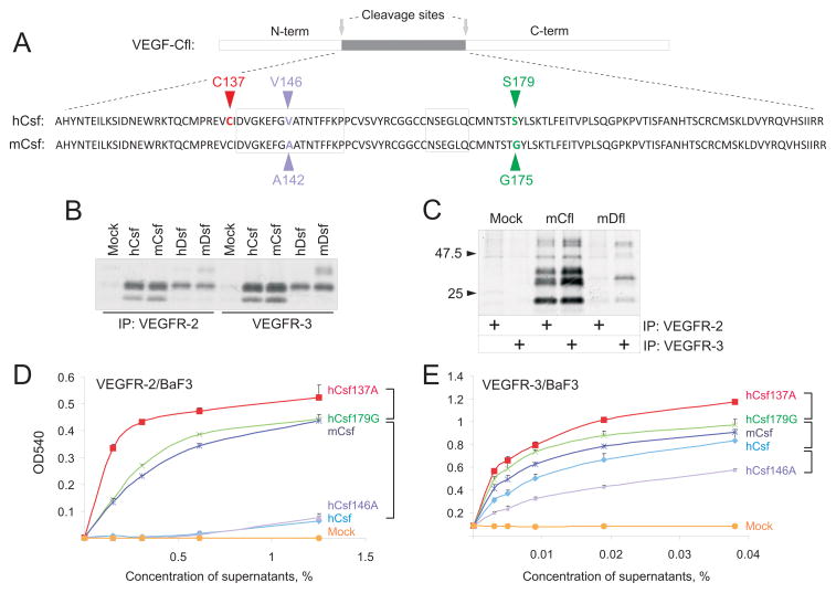
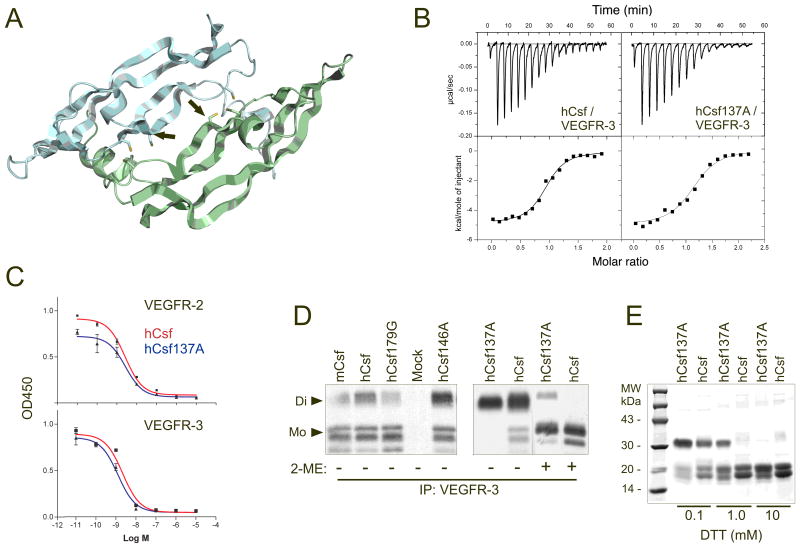
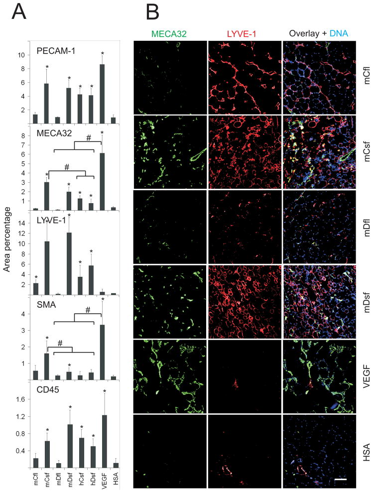
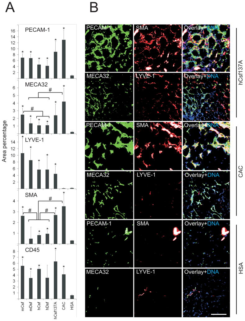
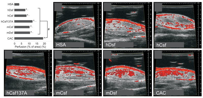
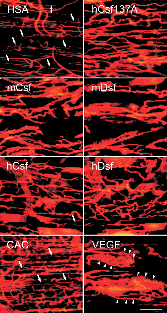
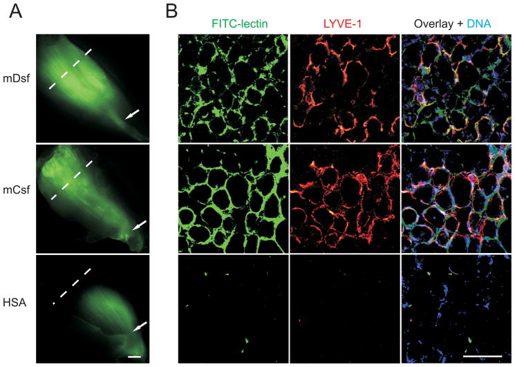
The therapeutic potential of vascular endothelial growth factor (VEGF)-C and VEGF-D in skeletal muscle has been of considerable interest as these factors have both angiogenic and lymphangiogenic activities. Previous studies have mainly used adenoviral gene delivery for short-term expression of VEGF-C and VEGF-D in pig, rabbit, and mouse skeletal muscles. Here we have used the activated mature forms of VEGF-C and VEGF-D expressed via recombinant adeno-associated virus (rAAV), which provides stable, long-lasting transgene expression in various tissues including skeletal muscle. Mouse tibialis anterior muscle was transduced with rAAV encoding human or mouse VEGF-C or VEGF-D. Two weeks later, immunohistochemical analysis showed increased numbers of both blood and lymph vessels, and Doppler ultrasound analysis indicated increased blood vessel perfusion. The lymphatic vessels further increased at the 4-week time point were functional, as shown by FITC-lectin uptake and transport. Furthermore, receptor activation and arteriogenic activity were increased by an alanine substitution mutant of human VEGF-C (C137A) having an increased dimer stability and by a chimeric CAC growth factor that contained the VEGF receptor-binding domain flanked by VEGF-C propeptides, but only the latter promoted significantly more blood vessel perfusion when compared to the other growth factors studied. We conclude that long-term expression of VEGF-C and VEGF-D in skeletal muscle results in the generation of new functional blood and lymphatic vessels. The therapeutic value of intramuscular lymph vessels in draining tissue edema and lymphedema can now be evaluated using this model system.


Figures

Figure 1
Figure 1. Biochemical characterisation of recombinant growth factors
A, Alignment of the human (h) and mouse (m) VEGF-Csf (Csf) sequences. The amino acid residue differences (V146/A142, S179/G175) are indicated in blue and green and the mutated residue (C137) in red. B, Soluble VEGFR-2 and VEGFR-3 precipitation of human and mouse factors followed by SDS-PAGE analysis under reducing conditions. Empty plasmid was used as the mock control. C, Coprecipitation of mouse full-length (VEGF-Cfl) and VEGF-Dfl with VEGFR-2 or VEGFR-3. D, MTT cell survival assay with VEGFR-2/BaF3 cells for the VEGF-Csf native and mutant molecules produced in transfected 293T cells. Mouse and human VEGF-Csf are denoted by mCsf and hCsf, respectively. V146A, S179G and C137A mutants of hVEGF-Csf are denoted as hCsf146A, hCsf179G, and hCsf137A, respectively. E, The same assay as in D, but utilizing VEGFR-3/BaF3 cells. In D and E, statistically significant (P<0.05) differences between the activities of certain proteins over range of studied concentrations are indicated by square brackets.
Figure 2
Figure 2. Biochemical properties of wild-type versus mutant VEGF-Csf and VEGF-Dsf
A, A 3-D computer model (SWISS-MODEL45) shows the putative location of the C137A mutation in the VEGF-Csf structure. The mutant residue (shown by red in Figure 1A) is marked with arrows. The two antiparallel polypeptide chains of hVEGF-Csf homodimer are shown in grey and green. The hVEGF-Csf model is based on the known crystal structure of VEGF amino terminal residues 8-109 and other VEGF family proteins,. B, Analysis of the VEGFR-3 binding affinities of human VEGF-Csf and its C137A mutant by isothermal titration calorimetry. The proteins were produced as described in Materials and Methods. C, ELISA-based competitive binding assay with VEGF-Csf and its C137A mutant. D, VEGFR-3 co-precipitation of [35S]-labelled human VEGF-Csf mutants or wild type proteins, which were obtained from the media of 293T cells transfected with the corresponding plasmids (Di - dimer, Mo - monomer). Mock represents the supernatant of 293T cells transfected with empty plasmid. E, SDS-PAGE of purified VEGF-Csf and its C137A mutant treated with varying concentrations of DTT. The proteins were stained with Coomassie Blue. Growth factor abbreviations are described in Figure 1 legend.
Figure 3
Figure 3. Blood vascular and lymphatic endothelial staining of the target muscles four weeks after gene transduction
Immunostaining for PECAM-1 (endothelial cells), MECA32 (blood vessel endothelial cells), LYVE-1 (lymphatic endothelial cells), SMA (smooth muscle cells and pericytes), and CD45 (leukocytes) frozen sections of the t.a. muscles of FVB/NJ mice. Full-length forms of mouse VEGF-Cfl and VEGF-Dfl were tested along with mouse and human VEGF-Csf and VEGF-Dsf. A, Quantification of the immunostaining was perfomed as described in Materials and Methods. * marks statistical significance at P<0.05, when compared to the HSA control. Square brackets with # mark statistical significance at P<0.05 between the indicated experimental groups. B, Representative images of mouse factor immunostaining are shown. The corresponding PECAM-1/SMA immunostaining patterns are shown in Online Figure III. Note that only mVEGF-Csf and VEGF165 induced significant recruitment of smooth muscle cells. Scale bars here and in all other figures are 100 μm, unless otherwise indicated.
Figure 4
Figure 4. Blood vascular, lymphatic endothelial, and smooth muscle staining two weeks after gene transduction
Double-immunostaining of the indicated antigens in frozen muscle sections from C57Bl/6J mice. A, Quantification of the immunostaining was performed as in Fig. 3. B, Representative images of hVEGF-Csf C137A-, CAC-, and HSA-treated muscle samples. Growth factor abbreviations are described in the Figure 1 legend. Note that while hVEGF-Csf C137A induces growth of both blood (MECA32-positive) and lymphatic (LYVE-1-positive) vessels, CAC induces only blood vessels. It should be noted that some of the inflammatory cells were also PECAM-1 positive.
Figure 5
Figure 5. Muscle perfusion two weeks after transduction of rAAV vectors
C57Bl/6J mice were separated into 7 groups, and 4 animals per group were injected with vectors encoding the indicated growth factors. Blood flow in t.a. muscle was quantified by Doppler ultrasound. Significance values were determined between the test groups and the negative control group (HSA). The bars indicate ± s.e.m.; * marks statistically significant differences to HSA control. # marks statistical significance between CAC and any other experimental group. Representative 2-D images from the scanning of the muscles transduced with the various vectors are indicated.
Figure 6
Figure 6. Comparison of perfusion staining of vessels induced by VEGF-C/-D, CAC and VEGF
Intramuscular blood vessels were stained by perfusion with the lipophilic dye DiI two weeks after rAAV transduction. Note that both VEGF-C and VEGF-D increase vessel density and size. In CAC treated muscles, vessel density is markedly increased and vessel morphology most similar to that of muscle injected with the control vector (HSA), whereas VEGF induced the formation of angioma-like structures (arrowheads). Capillary-sized vessels are indicated by arrows.
Figure 7
Figure 7. Mouse VEGF-Csf and VEGF-Dsf induce formation of functional lymphatic vessels
A, Lymphatic dye uptake and transfer were imaged 45 minutes after FITC-lectin injection into the distal (lower) part of t.a. muscles four weeks post-transduction with rAAVs encoding the indicated proteins. The stained sections were prepared from the regions indicated by the punctuated lines. Arrows indicate sites of FITC-lectin injection. The scale bar indicates 1 mm. B, FITC-lectin, nuclear (Hoechst 33258), and anti-LYVE-1 immunostaining were performed using muscle sections obtained from the muscle areas indicated in A.

References

    1. Takahashi H, Shibuya M. The vascular endothelial growth factor (VEGF)/VEGF receptor system and its role under physiological and pathological conditions. Clin Sci (Lond) 2005;109:227–241. - PubMed
    1. Yamazaki Y, Morita T. Molecular and functional diversity of vascular endothelial growth factors. Mol Divers. 2006;10:515–527. - PubMed
    1. Tirziu D, Simons M. Angiogenesis in the human heart: Gene and cell therapy. Angiogenesis. 2005;8:241–251. - PubMed
    1. Karpanen T, Alitalo K. Molecular biology and pathology of lymphangiogenesis. Annu Rev Pathol. 2008;3:367–397. - PubMed
    1. Makinen T, Veikkola T, Mustjoki S, Karpanen T, Catimel B, Nice EC, Wise L, Mercer A, Kowalski H, Kerjaschki D, Stacker SA, Achen MG, Alitalo K. Isolated lymphatic endothelial cells transduce growth, survival and migratory signals via the VEGF-C/D receptor VEGFR-3. EMBO J. 2001;20:4762–4773. - PMC - PubMed

Publication types

MeSH terms

Substances