. 2009 Jun 19;104(12):1390-8.
doi: 10.1161/CIRCRESAHA.108.192773. Epub 2009 May 14.

Antiarrhythmic drug-induced internalization of the atrial-specific k+ channel kv1.5

Affiliations

Antiarrhythmic drug-induced internalization of the atrial-specific k+ channel kv1.5

Sarah M Schumacher et al. Circ Res. .

Abstract

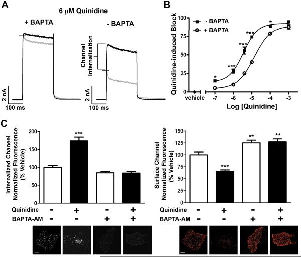
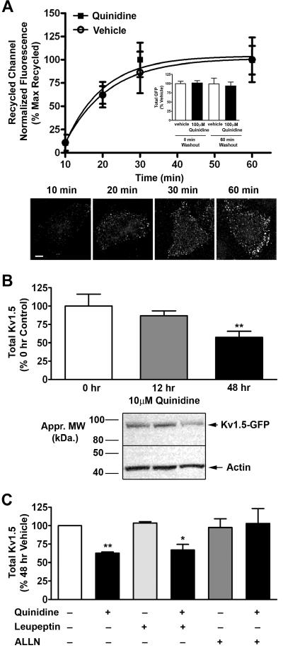
Conventional antiarrhythmic drugs target the ion permeability of channels, but increasing evidence suggests that functional ion channel density can also be modified pharmacologically. Kv1.5 mediates the ultrarapid potassium current (I(Kur)) that controls atrial action potential duration. Given the atrial-specific expression of Kv1.5 and its alterations in human atrial fibrillation, significant effort has been made to identify novel channel blockers. In this study, treatment of HL-1 atrial myocytes expressing Kv1.5-GFP with the class I antiarrhythmic agent quinidine resulted in a dose- and temperature-dependent internalization of Kv1.5, concomitant with channel block. This quinidine-induced channel internalization was confirmed in acutely dissociated neonatal myocytes. Channel internalization was subunit-dependent, activity-independent, stereospecific, and blocked by pharmacological disruption of the endocytic machinery. Pore block and channel internalization partially overlap in the structural requirements for drug binding. Surprisingly, quinidine-induced endocytosis was calcium-dependent and therefore unrecognized by previous biophysical studies focused on isolating channel-drug interactions. Importantly, whereas acute quinidine-induced internalization was reversible, chronic treatment led to channel degradation. Together, these data reveal a novel mechanism of antiarrhythmic drug action and highlight the possibility for new agents that selectively modulate the stability of channel protein in the membrane as an approach for treating cardiac arrhythmias.


Figures

Figure 1
Figure 1. Quinidine stimulates internalization of Kv1.5 in a dose- and temperature-dependent manner
(A) Representative images of Kv1.5-GFP overexpressed in HL-1 cells, showing total GFP signal (left), surface channel as detected by surface labeling with anti-GFP followed by goat anti-rabbit Alexa Fluor 405 (middle), and internalized channel detected by labeling with goat anti-rabbit Alexa Fluor 647 (right), at 0min (top), 10min at 37°C with vehicle (0.1% DMSO) (middle), and 10min at 37°C with 100 μmol/L quinidine (bottom). (B) Quantification of internalized (top) and surface (bottom) Kv1.5 following treatment with increasing concentrations of quinidine for 10min at 37°C. (C) Comparison of dose response for Kv1.5-GFP internalization following quinidine treatment for 10min at room temperature (25°C) and 37°C (EC50 = 900 nmol/L at 37°C). Scale bars = 10 μm. * indicates p < 0.05; *** indicates p < 0.001 as determined by oneway ANOVA with Tukey post-test.
Figure 2
Figure 2. Constitutive and quinidine-induced internalization occur in native mouse myocytes
(A) Representative images of acutely dissociated neonatal mouse myocytes transiently expressing Kv1.5-GFP by electroporation showing total GFP signal (left), surface channel (left middle), internalized channel (right middle), and anti-troponin (right) signal, at 0min (top), 10min at 37°C with vehicle (middle), and 10min at 37°C with 100 μmol/L quinidine (bottom). Scale bars = 10 μm. (B) Quantification of internalized (left) and surface (right) Kv1.5 following treatment with 100 μmol/L quinidine for 10min at 37°C. *** indicates p < 0.001 as determined by unpaired t-test.
Figure 3
Figure 3. Quinidine-induced internalization is subunit-dependent, activity-independent, and stereospecific
Dose-response for HL-1 cells expressing Kv4.2-GFP or Kv2.1-GFP (A) or Kv1.5-W472F (B) treated with increasing concentrations of quinidine for 10min at 37°C. ** indicates p < 0.01; *** indicates p < 0.001 as determined by one-way ANOVA with Tukey post-test. (C) Quantification of internalized Kv1.5 following treatment with 100 μmol/L quinidine or 200 μmol/L quinine, the diastereomer of quinidine, for 10min at 37°C. *** indicates p < 0.001 as determined by student's unpaired t-test.
Figure 4
Figure 4. Structural requirements for quinidine binding are partially conserved for pore block and channel internalization
(A) Dose-response for HL-1 cells expressing Kv1.5-GFP or the quinidine-insensitive Kv1.5-T480A mutant treated with increasing concentrations of quinidine for 10min at 37°C. (B) Quinidine-induced internalization for HL-1 cells expressing Kv1.5-GFP containing the T480A, I508A, L510A, or V512A mutation treated with 100 μmol/L quinidine for 10min at 37°C. (C) Dose-Response for HL-1 cells expressing Kv1.5-GFP or the Kv1.5-P532L mutant treated as described in (A). * indicates p < 0.05; ** indicates p < 0.01; *** indicates p < 0.001 as determined by one-way ANOVA.
Figure 5
Figure 5. Channel internalization is prevented by pharmacologic disruption of the endocytic machinery
(A) HL-1 cells expressing Kv1.5-GFP were treated for 1 hour with 80 μmol/L Dynasore at 37°C prior to surface-labeling with anti-GFP antibody. Quantification of internalized Kv1.5 following treatment with 100 μmol/L quinidine for 10min at 37°C in the continued presence of Dynasore. (B) Quantification of surface Kv1.5 for cells treated as described in (A). (C) Representative images of surface (red) and internalized (white) Kv1.5 in cells treated as described in (A). Scale bars = 10 μm. ** indicates p < 0.01; *** indicates p < 0.001 as determined by one-way ANOVA with Tukey post-test.
Figure 6
Figure 6. Quinidine-induced internalization occurs via a calcium-dependent mechanism
Whole-cell voltage clamp experiments were performed on HL-1 cells stably expressing Kv1.5-pHluorin. A single depolarizing pulse to +60mV was applied as described in methods. (A) Current traces are shown for a single cell prior to (black) and following (gray) 10min exposure to 6 μmol/L quinidine in the presence (left) or absence (right) of BAPTA in the pipette solution (n=10 cells). (B) Dose-response curve upon treatment with increasing concentrations of quinidine for 10min at room temperature in the presence or absence of BAPTA in the pipette solution (IC50 = 13 μmol/L + BAPTA and 3.5 μmol/L - BAPTA; n=5 cells; Hill slope = 1.042 + BAPTA and 1.135 - BAPTA). (C) HL-1 cells expressing Kv1.5-GFP were pretreated for 1 hour with 10 μmol/L BAPTA-AM before surface-labeling with anti-GFP antibody. Quantification of internalized (left) and surface (right) Kv1.5 following treatment with 100 μmol/L quinidine for 10min at 37°C in the continued presence of BAPTA-AM. Below each bar graph is a representative image for that condition. Scale bars = 10 μm. * indicates p < 0.05; ** indicates p < 0.01; *** indicates p < 0.001 as determined by one-way ANOVA with Tukey post-test.
Figure 7
Figure 7. Acute quinidine-induced internalization is reversible, whereas chronic treatment results in channel degradation
(A) Quantification of recycled Kv1.5 at 0, 10, 20, 30, and 60 min at 37°C post treatment with 100 μmol/L quinidine for 10 min at 37°C. Vehicle and quinidine treated data sets were each normalized to their own baseline (no washout) and maximum. Corresponding total GFP levels are provided for 0 and 60 min of recycling (inset). Below are representative images showing the increase in recycled Kv1.5-GFP with time, post quinidine treatment. Scale bar = 10 μm. (B) HL-1 cells stably expressing Kv1.5-pHluorin were treated with 10 μmol/L quinidine for 0, 12, or 48 hours at 37°C. Quantification of total Kv1.5 protein levels normalized to actin and control (48 hour DMSO) levels. Below is a representative image of a Western blot for 0, 12, and 48 hours of quinidine treatment. (C) HL-1 cells stably expressing Kv1.5-pHluorin were treated with vehicle, 10 μmol/L quinidine, 10 μmol/L leupeptin, 500 nmol/L ALLN, quinidine and leupeptin, or quinidine and ALLN for 48 hours at 37°C (63% or 67% decrease with 48 hr quinidine or quinidine and leupeptin (n = 8); no statistical decrease for quinidine and ALLN (n = 4)). Quantification was performed as in B. * indicates p < 0.05; ** indicates p < 0.01 as determined by one-way ANOVA with Tukey post-test.

References

    1. Lafuente-Lafuente C, Mouly S, Longas-Tejero MA, Bergmann JF. Antiarrhythmics for maintaining sinus rhythm after cardioversion of atrial fibrillation. Cochrane database of systematic reviews (Online) 2007:CD005049. - PubMed
    1. Lafuente-Lafuente C, Mouly S, Longas-Tejero MA, Mahe I, Bergmann JF. Antiarrhythmic drugs for maintaining sinus rhythm after cardioversion of atrial fibrillation: a systematic review of randomized controlled trials. Archives of internal medicine. 2006;166:719–728. - PubMed
    1. Nattel S, Carlsson L. Innovative approaches to anti-arrhythmic drug therapy. Nat Rev Drug Discov. 2006;5:1034–1049. - PubMed
    1. Waldo AL. A perspective on antiarrhythmic drug therapy to treat atrial fibrillation: there remains an unmet need. American heart journal. 2006;151:771–778. - PubMed
    1. Feng J, Wible B, Li GR, Wang Z, Nattel S. Antisense oligodeoxynucleotides directed against Kv1.5 mRNA specifically inhibit ultrarapid delayed rectifier K+ current in cultured adult human atrial myocytes. Circ Res. 1997;80:572–579. - PubMed

Publication types

MeSH terms