Comparative Study
. 1991 Nov 14;354(6349):159-61.
doi: 10.1038/354159a0.

Deletion of steroid 5 alpha-reductase 2 gene in male pseudohermaphroditism

Affiliations
Comparative Study

Deletion of steroid 5 alpha-reductase 2 gene in male pseudohermaphroditism

S Andersson et al. Nature. .

Abstract

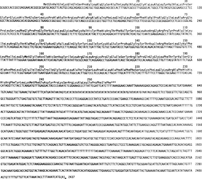
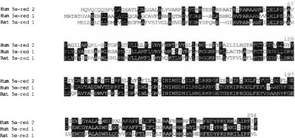
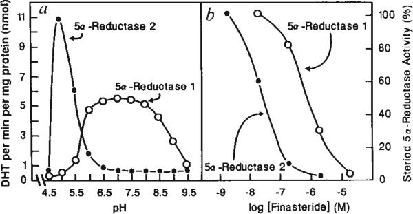
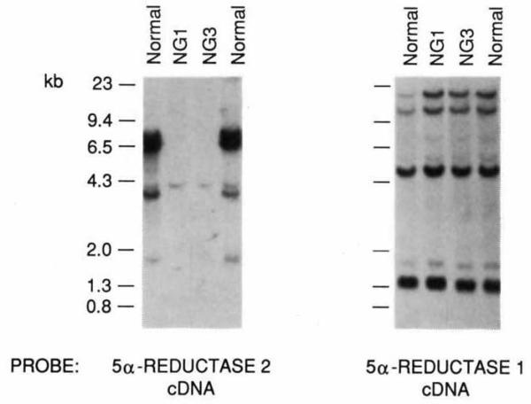
The conversion of testosterone into dihydrotestosterone by steroid 5 alpha-reductase is a key reaction in androgen action, and is essential both for the formation of the male phenotype during embryogenesis and for androgen-mediated growth of tissues such as the prostate. Single gene defects that impair this conversion lead to pseudohermaphroditism in which 46X,Y males have male internal urogenital tracts, but female external genitalia. We have described the isolation of a human 5 alpha-reductase complementary DNA from prostate. Subsequent cloning and genetic studies showed that this gene (designated 5 alpha-reductase 1) was normal in patients with 5 alpha-reductase deficiency. We report here the isolation of a second 5 alpha-reductase cDNA by expression cloning and the polymerase chain reaction. The biochemical and pharmacological properties of this cDNA-encoded enzyme (designated 5 alpha-reductase 2) are consistent with it being the major isozyme in genital tissue. A deletion in this gene is present in two related individuals with male pseudohermaphroditism caused by 5 alpha-reductase deficiency. These results verify the existence of at least two 5 alpha-reductases in man and provide insight into a fundamental hormone-mediated event in male sexual differentiation.


Figures

FIG. 1
FIG. 1
cDNA and amino-acid sequence of human 5α-reductase 2. Nucleotides are numbered at right. Amino acids are numbered above the protein sequence. METHODS. An oriented and size-fractionated cDNA library was constructed in a pCMV vector from human prostate poly(A)+ mRNA using a kit purchased from GIBCO-BRL. The cDNA was electroporated into Escherichia coli HB101 cells, and pools of about 104 independent cDNAs were grown overnight in 10 ml cultures of superbroth media. Plasmid DNA was prepared using Quiagentip 100 columns and 5 μg aliquots were transfected through a calcium phosphate procedure into 60 mm dishes of human embryonic kidney 293 cells (ATCC #CRL 1573). To enhance expression, 0.5 μg of a plasmid (pVA1) containing the adenovirus VAI gene was cotransfected with the pooled cDNAs. On day 2 of the transfection experiments, 14C-testosterone (120 d.p.m. nmol−1) was added to the medium at a final concentration of 1 μM, and conversion into dihydrotestosterone was determined 18 h later as described previously,. A pool expressing 5α-reductase enzyme activity was subsequently screened with a probe generated by a polymerase chain reaction in which two oligonucleotides, GA(A/G)TGGTG(T/C)T(T/A)(T/C)GCN(C/T)TNGC and TTIGG(A/G)TAITC(T/C)TC(A/G)AA(T/C)TT, encoding amino acids 205 to 211 and 243 to 249, respectively, of the human and rat 5α-reductase proteins,, were used to amplify random-primed cDNA synthesized from 0.4 μg total human prostate RNA. The reaction conditions were those of Strathmann et al., except that 30-s incubations at 94 °C, 40 °C, and 72 °C were used in place of those described. About one hybridization-positive was found per 104 colonies screened from the expressing pool. The DNA sequence of one such cDNA was determined on an Applied Biosystems Model 373 fluorescence sequencer after subcloning fragments into bacteriophage M13 vectors.
FIG. 2
FIG. 2
Alignment of 5α-reductase proteins. The amino-acid sequences in single-letter code of the human 5α-reductase 2, 5α-reductase 1 (ref. 4), and rat 5α-reductase proteins are aligned. Identities between two or more enzymes are boxed in black. Numbers above the sequences refer to the human 5α-reductase 2 protein.
FIG. 3
FIG. 3
Characterization of expressed 5α-reductase isozymes, a, pH optima. Enzyme activity was assayed at the indicated pH in cell extracts prepared from 293 cells transfected with the 5α-reductase 1 or 2 cDNA. Nontransfected cells express negligible amounts of enzyme activity, b, Inhibition by finasteride. Reductase enzyme activity obtained in extracts of transfected 293 cells in the absence of inhibitor is defined as 100%. In the presence of Increasing concentrations of finasteride, progressively less 5α-reductase enzyme activity was detected. METHODS. An expression plasmid containing the 5α-reductase 2 cDNA (Fig. 1) or the 5α-reductase 1 cDNA (ref. 4) was transfected into 293 cells as described in the legend to Fig. 1. In a, 48 h after transfection, cell extracts were prepared as described previously and 10 μg cell protein were assayed for 5α-reductase enzyme activity in 0.1 M Tris-citrate buffers at the indicated pH with 10 μM 14C-testosterone (120 d.p.m. pmol−1) as substrate and 10 mM NADPH as cofactor. In b, 5 μg of transfected cell protein were assayed in duplicate for 5α-reductase activity in the presence of the indicated concentration of finasteride (MK-906) (17β-(N-t-butyl)carbamoyl-4-aza-5α-androst-l-ene-3-one, a kind gift of G. Rasmusson, Merck, Sharp and Dohme), 4 μM 14C-testosterone (120 d.p.m. pmol−1) and 10 mM NADPH. In both panels, conversion into dihydrotesterone was determined after 10-min incubations by thin layer chromatography as described previously. Protein concentrations in cell extracts were measured by Lowry assay.
FIG. 4
FIG. 4
Deletion of 5α-reductase 2 gene in subjects with 5α-reductase deficiency. DNA isolated from two normal individuals and two related 5α-reductase deficiency subjects from a geographically isolated tribe in the Highlands of Papua New Guinea was screened by Southern blotting using the indicated 5α-reductase cDNA probes. The normal DNA analysed in the most left-hand lane was isolated from an individual from the same New Guinea tribe as the NG1 and NG3 subjects, whereas the normal DNA analysed in the right-hand lane was isolated from a caucasian American. The filter was screened with the 5α-reductase 2 cDNA probe first, and then stripped and reprobed with the 5α-reductase 1 cDNA probe. A deletion of all but a weakly hybridizing fragment of about 4.5 kilobases in the DNA of the affected NG1 and NG3 individuals is apparent from the autoradiogram obtained with the 5α-reductase 2 probe. All individuals have a normal 5α-reductase 1 gene. METHODS. Genomic DNA was isolated from peripheral blood samples from the indicated individuals and prepared and analysed by high-stringency Southern blotting as described previously. Aliquots of DNA (20 μg) were digested with Hindlll before electrophoresis on agarose gels. Three single-stranded 32P-labelled probes spanning the coding region of the 5α-reductase 2 cDNA shown in Fig. 1 were prepared as described by Church and Gilbert and used in hybridization. After autoradiography for 5 days at −70 °C, the filter was stripped and reprobed with a random hexanucleotide 32P-labelled probe corresponding to the full-length 5α-reductase 1 cDNA (ref. 4).

References

    1. Wilson. J. D. Harvey Led. 1985;79:145–172. - PubMed
    1. Wilson JD. Endocrinology. 1975;25:491–508.
    1. Griffin JE, Wilson JD. In: The Metabolic Basis of Inherited Disease. Scriver CR, Beaudet AL, Sly WS, Valle D, editors. McGraw-Hill; New York: 1989. pp. 1919–1944.
    1. Andersson S, Russell DW. Proc. natn. Acad. Sci. U.S.A. 1990;87:3640–3644. - PMC - PubMed
    1. Andersson S, Davis D, Dahlback, Jornvail H, Russell DW. J. biol. Chem. 1989;264:8222–8229. - PubMed

Publication types

Substances