. 2009 May 29;137(5):873-86.
doi: 10.1016/j.cell.2009.03.046. Epub 2009 May 14.

DEPTOR is an mTOR inhibitor frequently overexpressed in multiple myeloma cells and required for their survival

Affiliations

DEPTOR is an mTOR inhibitor frequently overexpressed in multiple myeloma cells and required for their survival

Timothy R Peterson et al. Cell. .

Abstract

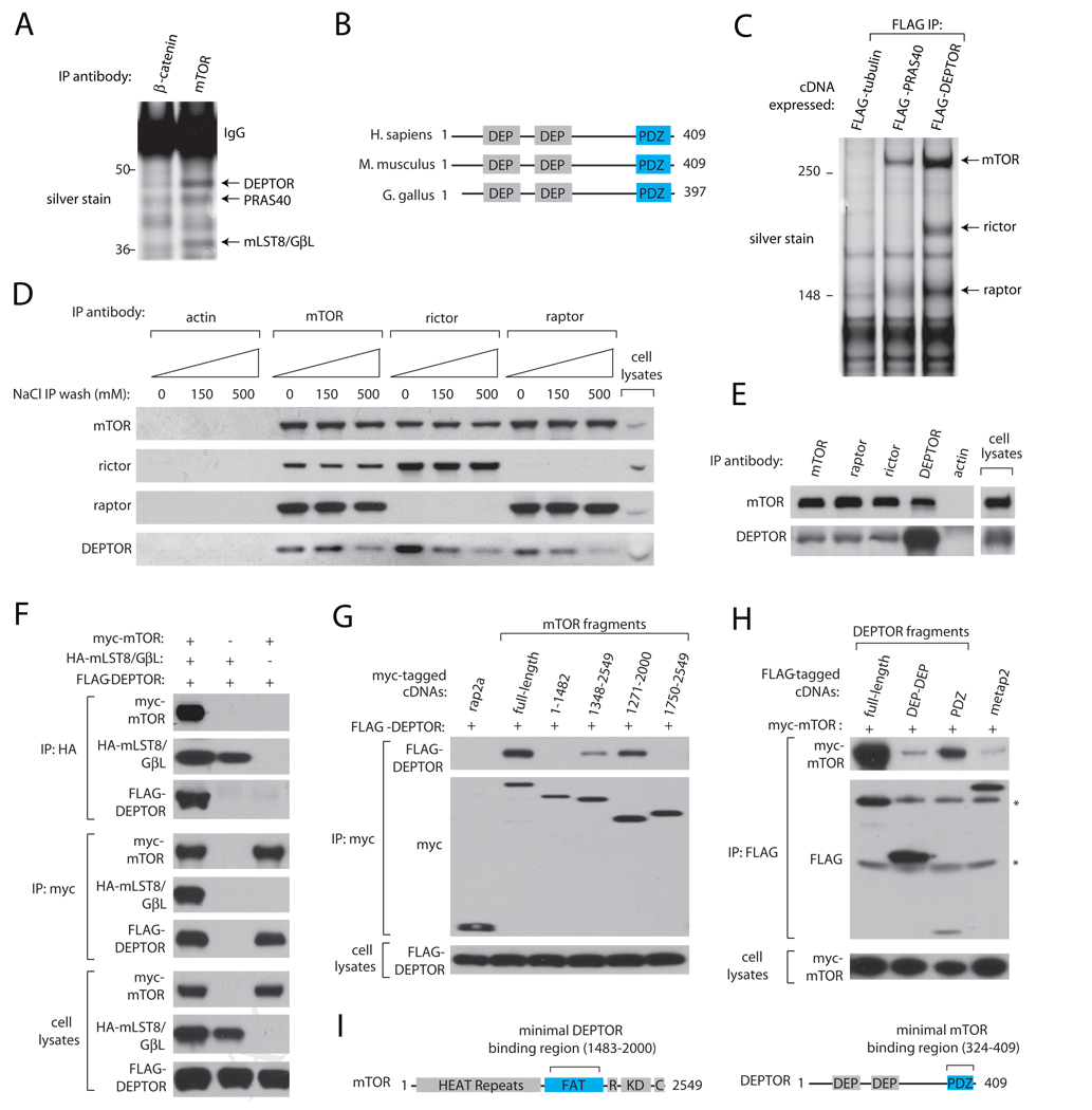
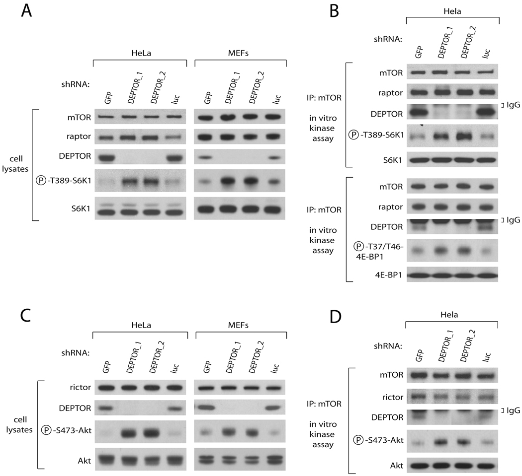
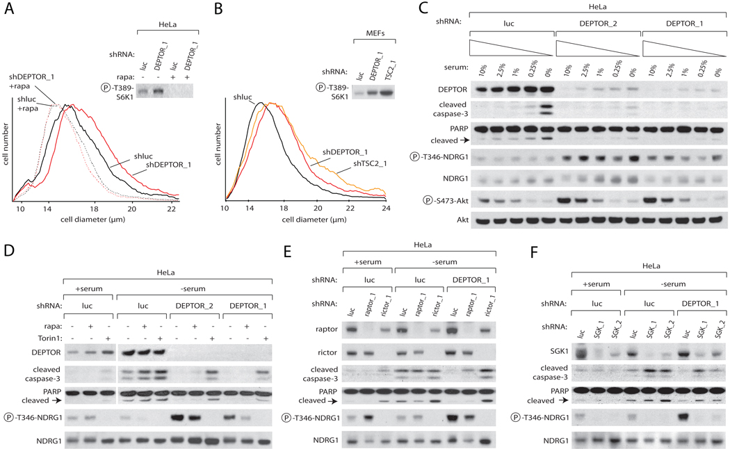
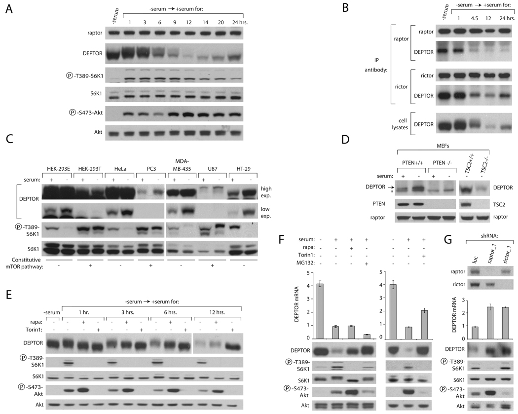
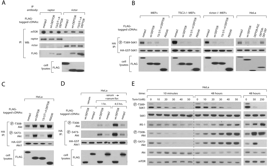
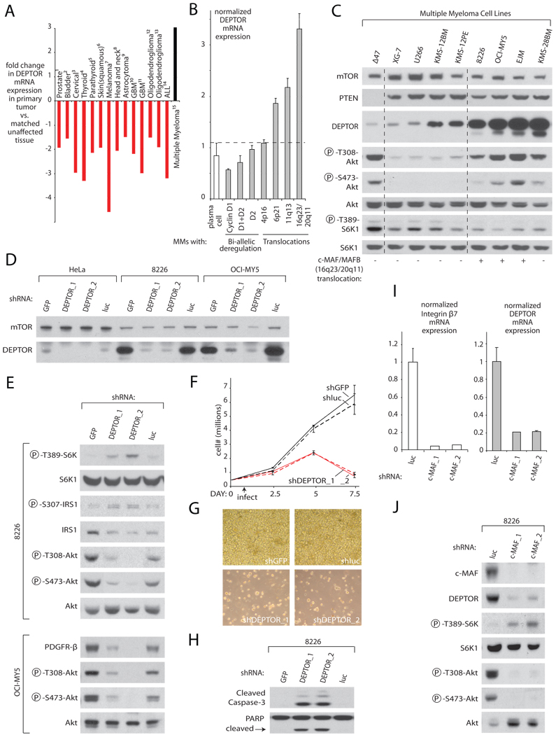
The mTORC1 and mTORC2 pathways regulate cell growth, proliferation, and survival. We identify DEPTOR as an mTOR-interacting protein whose expression is negatively regulated by mTORC1 and mTORC2. Loss of DEPTOR activates S6K1, Akt, and SGK1, promotes cell growth and survival, and activates mTORC1 and mTORC2 kinase activities. DEPTOR overexpression suppresses S6K1 but, by relieving feedback inhibition from mTORC1 to PI3K signaling, activates Akt. Consistent with many human cancers having activated mTORC1 and mTORC2 pathways, DEPTOR expression is low in most cancers. Surprisingly, DEPTOR is highly overexpressed in a subset of multiple myelomas harboring cyclin D1/D3 or c-MAF/MAFB translocations. In these cells, high DEPTOR expression is necessary to maintain PI3K and Akt activation and a reduction in DEPTOR levels leads to apoptosis. Thus, we identify a novel mTOR-interacting protein whose deregulated overexpression in multiple myeloma cells represents a mechanism for activating PI3K/Akt signaling and promoting cell survival.


Figures

Figure 1
Figure 1. DEPTOR is an mTOR-Interacting Protein
(A) Silver stain of SDS-PAGE analysis of mTOR immunoprecipitates prepared from HEK-293E cells. (B) Schematic representation of structural features of human DEPTOR and its mouse and chicken orthologues. (C) Endogenous mTOR, raptor, and rictor co-immunoprecipitate with epitope-tagged DEPTOR. FLAG immunoprecipitates from HEK-293E cells expressing FLAG-DEPTOR, FLAG-PRAS40, or FLAG-tubulin were analyzed by SDS-PAGE followed by silver staining. (D) Interaction of endogenous DEPTOR with endogenous mTORC1 and mTORC2 is sensitive to high salt-containing buffers. mTOR, raptor, rictor, or actin immunoprecipitates were prepared from HEK-293E cells, washed with buffers containing indicated amounts of NaCl and analyzed by SDS-PAGE followed by immunoblotting for indicated proteins. (E) Endogenous DEPTOR co-immunoprecipitates endogenous mTOR. DEPTOR immunoprecipitates were prepared from HeLa cells, washed in a buffer containing 150 mM NaCl, and analyzed as in (D). (F) DEPTOR interacts with mTOR and not mLST8/GβL. Indicated cDNAs in expression vectors were co-expressed in HEK-293T cells, cell lysates prepared, and used to prepare anti-HA immunoprecipitates and anti-myc immunoprecipitates. Both immunoprecipitates and cell lysates were analyzed as in (D). (G) Myc-tagged mTOR, its indicated fragments, or rap2a were co-expressed in HEK-293T cells with FLAG-DEPTOR, and anti-myc immunoprecipitates were analyzed as in (D). (H) The PDZ domain of DEPTOR interacts with mTOR. FLAG-tagged DEPTOR, its fragments, or metap2 were co-expressed in HEK-293T cells with myc-mTOR, and anti-FLAG immunoprecipitates were analyzed as in (D). Asterisks (*) indicate non-specific bands. (I) Schematic indicating the regions of mTOR and DEPTOR that mediate their interaction. The abbreviations for and residues that define known mTOR domains are: HEAT Repeats, 1–1382; FAT, 1383–2014; R (FKBP12-Rapamycin Binding), 2015–2114; KD (PI3K-like Kinase Domain), 2115–2431; C (FATC), 2432–2549.
Figure 2
Figure 2. DEPTOR Depletion in Cells Activates mTORC1 and mTORC2 signaling and In Vitro Kinase Activities
(A) Knockdown of DEPTOR activates the mTORC1-S6K1 pathway. HeLa cells or p53−/− MEFs were infected with lentiviruses expressing shRNAs targeting the indicated genes. Cell lysates were analyzed by immunoblotting for the levels of the indicated proteins and phosphorylation states. (B) Knockdown of DEPTOR activates mTORC1 kinase activity towards S6K1 and 4E-BP1. HeLa cells were infected with lentiviruses expressing shRNAs targeting the indicated genes. mTOR immunoprecipitates were prepared from cell lysates (1 mg total protein) and analyzed for mTORC1 kinase activity toward S6K1 and 4E-BP1 and for levels of mTOR, raptor, and DEPTOR. (C) Knockdown of DEPTOR activates the mTORC2-Akt pathway. The experiment was performed as in (A) except that Akt phosphorylation and levels were measured. (D) Knockdown of DEPTOR activates mTORC2 kinase activity towards Akt1. The experiment was performed as in (B) except that mTORC2 activity towards Akt was measured.
Figure 3
Figure 3. DEPTOR Depletion Increases Cell Size and Protects Cells from Apoptosis Induced by Serum Deprivation
(A) Knockdown of DEPTOR increases cell size in an mTORC1-dependent fashion. Cell size distributions (graphs) and S6K1 phosphorylation (immunoblot) are shown for HeLa cells stably expressing shRNAs targeting DEPTOR or luciferase. Where indicated, cells were pre-treated with 100 nM rapamycin or vehicle before infection. 72 hours after infection, cell size was measured using a Coulter counter. (B) A knockdown of DEPTOR has similar effects on cells size as a TSC2 knockdown. p53 null MEFs were infected with shRNAs and cell size measured as in (A). (C) DEPTOR knockdown protects against apoptosis induced by serum withdrawal. HeLa cells expressing shRNAs targeting luciferase or DEPTOR were grown in media containing the indicated concentrations of serum for 30 hours. Cell lysates were then analyzed by immunoblotting for the levels of the indicated proteins and phosphorylation states. (D) mTOR inhibition re-sensitizes DEPTOR knockdown cells to apoptosis induced by serum withdrawal. HeLa cells expressing shRNAs targeting luciferase or DEPTOR were serum deprived for 30 hours in the presence of 100 nM rapamycin, 250 nM Torin1, or vehicle. Cell lysates were then analyzed as in (C). (E) Rictor but not raptor inhibition re-sensitizes DEPTOR knockdown cells to apoptosis induced by serum withdrawal. HeLa cells co-expressing shRNAs targeting luciferase or DEPTOR along with shRNAs targeting luciferase or rictor or raptor were serum deprived for 30 hours. Cell lysates were then analyzed as in (C). (F) SGK1 inhibition re-sensitizes DEPTOR knockdown cells to apoptosis induced by serum withdrawal. HeLa cells co-expressing shRNAs targeting luciferase or DEPTOR along with shRNAs targeting luciferase or SGK1 were analyzed as in (C).
Figure 4
Figure 4. mTOR Negatively Regulates DEPTOR Protein and mRNA Expression
(A) Serum starvation and stimulation regulates DEPTOR protein expression. HeLa cells were serum starved for 30 hours and stimulated with serum (10% final concentration) for the indicated times. Cell lysates were analyzed by immunoblotting for the levels of indicated proteins and phosphorylation states. (B) DEPTOR amounts in mTORC1 and mTORC2 correlate with its expression within cells. HeLa cells were serum starved for 30 hours and stimulated with serum (10% final concentration) for the indicated times. Cell lysates and raptor and rictor immunoprecipitates were analyzed by immunoblotting for indicated proteins. (C) DEPTOR protein expression is downregulated in cancer cell lines with constitutive mTOR signaling. Cell lines were seeded at equal density and grown in the presence or absence of serum for 30 hours and cell lysates analyzed as in (A). (D) DEPTOR protein expression is downregulated in PTEN null or TSC2 null MEFs. Indicated MEF lines were seeded at equal density and grown in the presence or absence of serum for 30 hours and cell lysates analyzed for levels of DEPTOR, PTEN, TSC2, and raptor. An asterisk (*) indicates a non-specific band. (E) Rapamycin treatment partially and Torin1 treatment fully rescues the serum-induced decrease in DEPTOR protein. HeLa cells were serum-deprived for 30 hours, and then stimulated with serum in the presence of 100 nM rapamycin, 250 nM Torin1, or vehicle for the specified times. Cell lysates were analyzed as in (A). (F) Regulation of DEPTOR protein and mRNA expression. HeLa cells were serum-deprived for 30 hours, and then stimulated with serum in the presence of 100 nM rapamycin, 10 uM of MG132, 250 nM Torin1, or vehicle for the specified times. Cell lysates were analyzed by immunoblotting for the levels of indicated proteins and phosphorylation states. DEPTOR mRNA was measured by qRT-PCR and normalized to GADPH mRNA levels. Error bars indicate standard deviation for n=3. (G) Reductions in raptor or rictor expression increase DEPTOR protein and mRNA levels. Five days after transductions with raptor or rictor shRNA-expressing lentiviruses, HeLa cells were lysed and analyzed as in (A). DEPTOR mRNA was prepared and analyzed as in (F).
Figure 5
Figure 5. DEPTOR Overexpression Inhibits mTORC1 but Activates PI3K/Akt Signaling
(A) The 13×S/T->A DEPTOR mutant binds mTORC1 and mTORC2 more tightly than wild-type DEPTOR. HEK-293T cells were transfected with 500 ng of expression vectors encoding the indicated FLAG-tagged proteins for 48 hours. Raptor or rictor immunoprecipitates were prepared from cell lysates, washed with buffers containing 150 mM NaCl, and analyzed along with cell lysates by immunoblotting for indicated proteins. (B) Overexpression of DEPTOR inhibits T389 phosphorylation of S6K1. HeLa cells or the indicated p53−/− MEFS were cotransfected with expression plasmids encoding HA-GST-S6K1 as well as the indicated FLAG-tagged proteins. Cell lysates were prepared 24 hours after transfection and were analyzed by immunoblotting for the levels of the indicated proteins and phosphorylation states. (C) Overexpression of DEPTOR activates T308 and S473 Akt1 phosphorylation. HeLa cells were cotransfected with expression plasmids encoding HA-GST-Akt1 as well as the indicated FLAG-tagged proteins. Cell lysates were analyzed as in (B). (D) Overexpression of DEPTOR activates T308 and S473 Akt1 phosphorylation only in serum-replete cells. HeLa cells were transfected as in (B), serum starved for 24 hours and stimulated for the indicated times and cell lysates were analyzed as in (B). (E) Partial inhibition of mTOR with Torin1 inhibits S6K1 phosphorylation but activates PI3K/mTORC2/Akt signaling. HeLa cells were treated with the specified concentrations of Torin1 or vehicle for either 10 minutes or 48 hours and cell lysates were analyzed as in (B).
Figure 6
Figure 6. DEPTOR is Overexpressed in a Subset of Multiple Myeloma Cells and in these Cells Activates PI3K/Akt signaling and Promotes Cell Survival
(A) DEPTOR mRNA expression is downregulated in most cancers, yet is up-regulated in Multiple Myelomas. Relative DEPTOR mRNA expression in various cancer types versus matched unaffected tissue was collated from publicly available microarray studies. For all studies shown, p<0.05 for DEPTOR mRNA downregulation or upregulation by one-tailed T-test. (B) DEPTOR mRNA is mostly highly expressed in the subset of Multiple Myelomas which possess Cyclin D1/D3 or c-MAF/MAFB translocations. DEPTOR mRNA levels were normalized by the summed mean values of the housekeeping genes GAPDH, β-actin, and FIP (7 total probe-sets). Error bars indicate standard error for n=7. The differences in mean DEPTOR mRNA levels between plasma cells and MMs with Cyclin D1, Cyclin D3, or c-MAF/MAFB translocations are significant to at least p<0.005. (C) Human Multiple Myeloma cell lines with high DEPTOR protein expression have activated PI3K and suppressed mTORC1 signaling. Cell lysates were analyzed by immunoblotting for the levels of the indicated proteins and phosphorylation states. Cell lines with known translocations of c-MAF or MAFB are indicated. (D) RNAi-mediated knockdown of DEPTOR in 8226 and OCI-MY5 cells reduces DEPTOR expression to levels similar to those in HeLa cells. Five days after transductions with shRNA-expressing lentiviruses, cells were lysed and as in (C). (E) DEPTOR suppression in 8226 and OCI-MY5 cells activates mTORC1 signaling, inhibits IRS-1 or PDGFR-β expression, and reduces PI3K/mTORC2/Akt signaling. Five days after transduction with shRNA-expressing lentiviruses, cells were lysed and analyzed as in (C). (F) DEPTOR suppression markedly decreases 8226 cell number. Cell number was measured at indicated time points with a Coulter Counter. Error bars indicate mean ± standard deviation for n=3 samples per time point. (G) Representative 20× light microscopy images of 8226 cells expressing shRNAs targeting DEPTOR, GFP, or luciferase at day 8 post-infection. (H) DEPTOR suppression causes apoptosis. Three days after transduction with shRNA-expressing lentiviruses, 8226 cells were lysed and analyzed as in (C). (I) A reduction in c-MAF expression decreases DEPTOR mRNA levels. Five days after transduction of 8226 cells with lentiviruses expressing the indicated shRNAs, DEPTOR and Integrin β7 mRNA levels were measured by qRT-PCR and normalized to GADPH mRNA levels. Error bars indicate standard deviation for n=3. (J) A c-MAF knockdown activates mTORC1 and downregulates PI3K signaling. Five days after transductions with shRNA-expressing lentiviruses, lysates of 8226 cells were analyzed as in (C).

References

    1. Alessi DR. Discovery of PDK1, one of the missing links in insulin signal transduction. Colworth Medal Lecture. Biochem Soc Trans. 2001;29:1–14. - PubMed
    1. Ballon DR, Flanary PL, Gladue DP, Konopka JB, Dohlman HG, Thorner J. DEP-domain-mediated regulation of GPCR signaling responses. Cell. 2006;126:1079–1093. - PubMed
    1. Bergsagel PL, Kuehl WM, Zhan F, Sawyer J, Barlogie B, Shaughnessy J., Jr Cyclin D dysregulation: an early and unifying pathogenic event in multiple myeloma. Blood. 2005;106:296–303. Epub 2005 Mar 2008. - PMC - PubMed
    1. Bliskovsky V, Ramsay ES, Scott J, DuBois W, Shi W, Zhang S, Qian X, Lowy DR, Mock BA. Frap, FKBP12 rapamycin-associated protein, is a candidate gene for the plasmacytoma resistance locus Pctr2 and can act as a tumor suppressor gene. Proc Natl Acad Sci U S A. 2003;100:14982–14987. Epub 12003 Nov 14921. - PMC - PubMed
    1. Brunn GJ, Hudson CC, Sekulic A, Williams JM, Hosoi H, Houghton PJ, Lawrence JC, Jr, Abraham RT. Phosphorylation of the translational repressor PHAS-I by the mammalian target of rapamycin. Science. 1997;277:99–101. - PubMed

Publication types