

Review
doi: 10.1016/j.semcdb.2009.04.001.
Epub 2009 Apr 10.
Signals from the edges: the cortical hem and antihem in telencephalic development
Affiliations
- PMID: 19446478
- PMCID: PMC2791850
- DOI: 10.1016/j.semcdb.2009.04.001
Item in Clipboard
Review
Signals from the edges: the cortical hem and antihem in telencephalic development
Semin Cell Dev Biol.
2009 Aug.
Abstract
The early cortical primordium develops from a sheet of neuroepithelium that is flanked by distinct signaling centers. Of these, the hem and the antihem are positioned as longitudinal stripes, running rostro-caudally along the medial and lateral faces, respectively, of each telencepahlic hemisphere. In this review we examine the similarities and differences in how these two signaling centers arise, their roles in patterning adjacent tissues, and the cells and structures they contribute to. Since both the hem and the antihem have been identified across many vertebrate phyla, they appear to be part of an evolutionary conserved set of mechanisms that play fundamental roles in forebrain development.
Figures
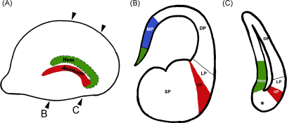
The positions of the hem and the antihem in the dorsal telencephalon. (A) A schematic of an E12.5 telencephalic hemisphere viewed from the lateral face, showing the antihem (red). The hem is schematized on the medial face (green). Rostral is to the left. (B) and (C) are schematics representing mid-level and caudal sections of such a hemisphere, showing the medial pallium (MP) which includes the hem (green) and the hippocampal primordium (blue), the dorsal pallium (DP), lateral pallium (LP), ventral pallium in red (VP/antihem), and subpallium (SP). Asterisk denotes DP tissue present between the hem and antihem at extreme caudal levels, which is the source of the amygdaloid nucleus nLOT2. Modified from Remedios et al. [www.nature.com/neuro/index.html ]. (For interpretation of the references to color in this figure legend, the reader is referred to the web version of the article.)
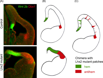
Lhx2 suppresses hem and antihem fate in a position-dependent manner. (A) Hem marker Wnt2b (green) and antihem marker Dbx1 (red) reveal these structures separated by the cortical neuroepithelium in a control E12.5 brain. In the Lhx2 mutant both the hem and the antihem are expanded and there is no intervening cortical neuroepithelium. (B) Schematics representing these data. (C) In a chimeric brain with Lhx2 null clusters scattered amidst wild-type neuroepithelium, the medial null patches take on hem identity whereas lateral patches differentiate into antihem. From Mangale et al. [www.sciencemag.org ]. (For interpretation of the references to color in this figure legend, the reader is referred to the web version of the article.)
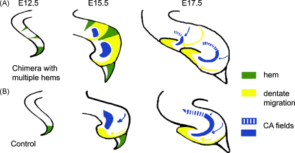
Ectopic hem induces and organizes multiple hippocampal fields. (A) and (B) are schematic representations of chimeric and control brains, respectively. The normal hem is seen at the medial extreme of the E12.5 telencephalic neuroepithelium. In the chimeras, Lhx2 mutant clusters form ectopic patches of hem in the medial telencephalon. By E15.5, control brains display markers for the hippocampal CA fields as well as for dentate granule cells. Both cell types originate in neuroepithelium and migrate away (blue and yellow arrows) to form the characteristic morphology of the Ammon's horn and the dentate gyrus by E17.5. In chimeric brains, CA and dentate cells are induced in appropriate spatial order adjacent to each ectopic hem, with the dentate granule cells immediately adjacent to the hem. By E17.5, the chimeras have assembled distinct dentate gyri and CA fields forming a double hippocampus. Modified from [www.sciencemag.org ]. (For interpretation of the references to color in this figure legend, the reader is referred to the web version of the article.)
References
-
- Remedios R., Huilgol D., Saha B., Hari P., Bhatnagar L., Kowalczyk T. A stream of cells migrating from the caudal telencephalon reveals a link between the amygdala and neocortex. Nat Neurosci. 2007;10:1141–1150. - PubMed
-
- Puelles L., Kuwana E., Puelles E., Bulfone A., Shimamura K., Keleher J. Pallial and subpallial derivatives in the embryonic chick and mouse telencephalon, traced by the expression of the genes Dlx-2, Emx-1, Nkx-2.1, Pax-6, and Tbr-1. J Comp Neurol. 2000;424:409–438. - PubMed
-
- Medina L., Legaz I., González G., De Castro F., Rubenstein J.L., Puelles L. Expression of Dbx1, Neurogenin 2, Semaphorin 5A, Cadherin 8, and Emx1 distinguish ventral and lateral pallial histogenetic divisions in the developing mouse claustroamygdaloid complex. J Comp Neurol. 2004;474:504–523. - PubMed
Publication types
MeSH terms
Substances
Grants and funding
LinkOut - more resources
Full Text Sources
