

Human HMGB1 directly facilitates interactions between nucleotide excision repair proteins on triplex-directed psoralen interstrand crosslinks
- PMID: 19446504
- PMCID: PMC2700216
- DOI: 10.1016/j.dnarep.2009.04.001
Human HMGB1 directly facilitates interactions between nucleotide excision repair proteins on triplex-directed psoralen interstrand crosslinks
Abstract
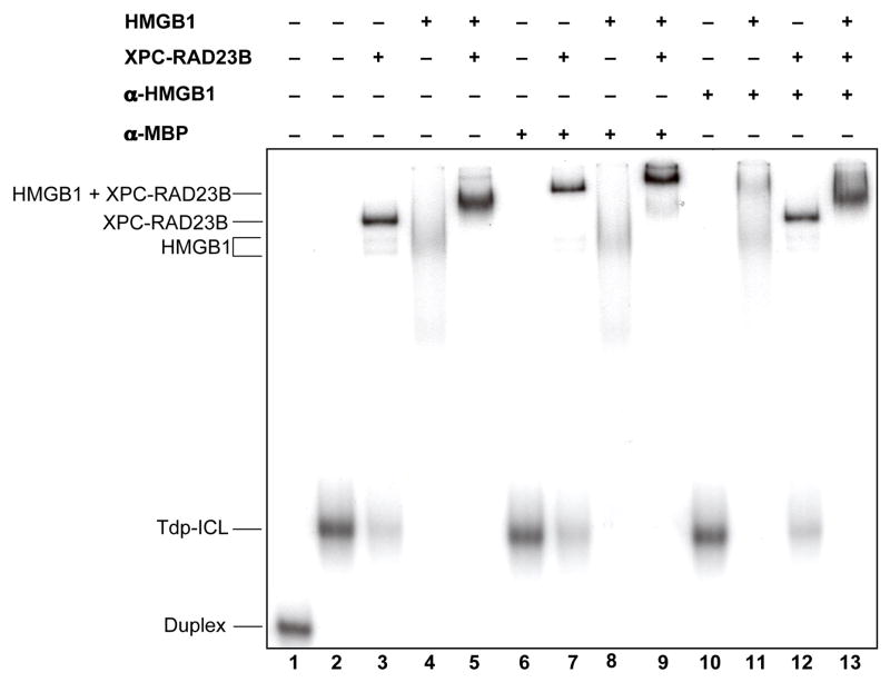
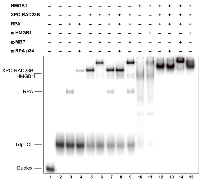
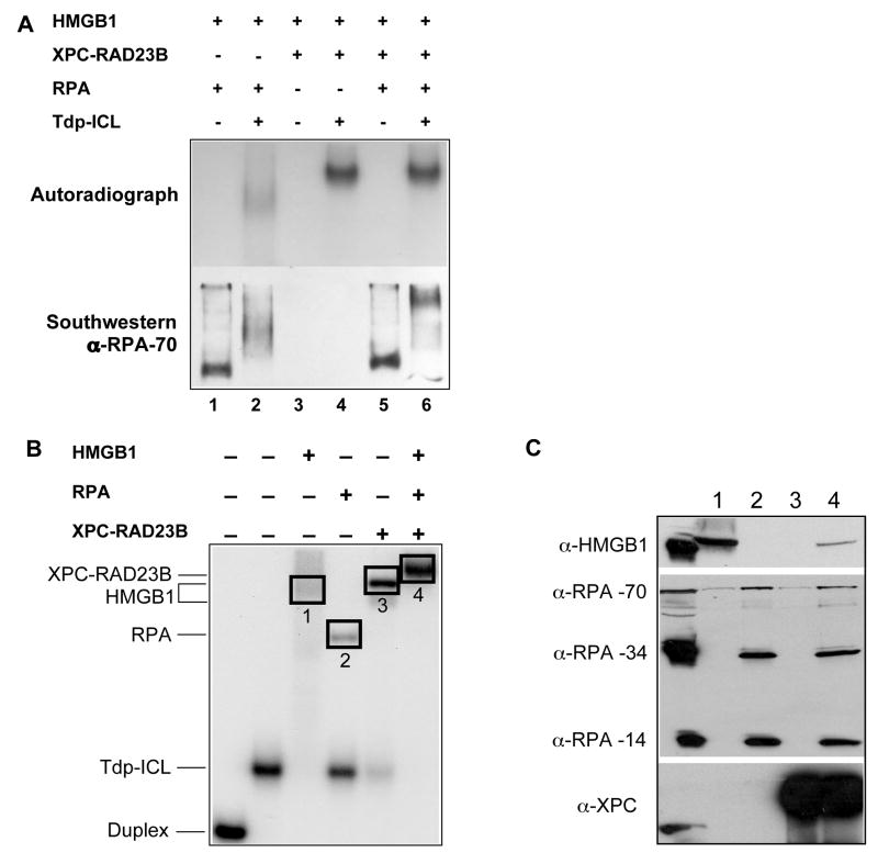
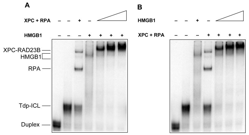
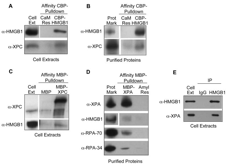
Psoralen is a chemotherapeutic agent that acts by producing DNA interstrand crosslinks (ICLs), which are especially cytotoxic and mutagenic because their complex chemical nature makes them difficult to repair. Proteins from multiple repair pathways, including nucleotide excision repair (NER), are involved in their removal in mammalian cells, but the exact nature of their repair is poorly understood. We have shown previously that HMGB1, a protein involved in chromatin structure, transcriptional regulation, and inflammation, can bind cooperatively to triplex-directed psoralen ICLs with RPA, and that mammalian cells lacking HMGB1 are hypersensitive to psoralen ICLs. However, whether this effect is mediated by a role for HMGB1 in DNA damage recognition is still unknown. Given HMGB1's ability to bind to damaged DNA and its interaction with the RPA protein, we hypothesized that HMGB1 works together with the NER damage recognition proteins to aid in the removal of ICLs. We show here that HMGB1 is capable of binding to triplex-directed psoralen ICLs with the dedicated NER damage recognition complex XPC-RAD23B, as well as XPA-RPA, and that they form a higher-order complex on these lesions. In addition, we demonstrate that HMGB1 interacts with XPC-RAD23B and XPA in the absence of DNA. These findings directly demonstrate interactions between HMGB1 and the NER damage recognition proteins, and suggest that HMGB1 may affect ICL repair by enhancing the interactions between NER damage recognition factors.
Figures
Similar articles
-
Human XPC-hHR23B interacts with XPA-RPA in the recognition of triplex-directed psoralen DNA interstrand crosslinks.Nucleic Acids Res. 2005 May 24;33(9):2993-3001. doi: 10.1093/nar/gki610. Print 2005. Nucleic Acids Res. 2005. PMID: 15914671 Free PMC article.
-
Mismatch repair and nucleotide excision repair proteins cooperate in the recognition of DNA interstrand crosslinks.Nucleic Acids Res. 2009 Jul;37(13):4420-9. doi: 10.1093/nar/gkp399. Epub 2009 May 25. Nucleic Acids Res. 2009. PMID: 19468048 Free PMC article.
-
HMGB1 interacts with XPA to facilitate the processing of DNA interstrand crosslinks in human cells.Nucleic Acids Res. 2016 Feb 18;44(3):1151-60. doi: 10.1093/nar/gkv1183. Epub 2015 Nov 17. Nucleic Acids Res. 2016. PMID: 26578599 Free PMC article.
-
Molecular basis for damage recognition and verification by XPC-RAD23B and TFIIH in nucleotide excision repair.DNA Repair (Amst). 2018 Nov;71:33-42. doi: 10.1016/j.dnarep.2018.08.005. Epub 2018 Aug 23. DNA Repair (Amst). 2018. PMID: 30174301 Free PMC article. Review.
-
Critical DNA damage recognition functions of XPC-hHR23B and XPA-RPA in nucleotide excision repair.Mol Carcinog. 2003 Sep;38(1):1-13. doi: 10.1002/mc.10143. Mol Carcinog. 2003. PMID: 12949838 Review.
Cited by
-
HMGB1: roles in base excision repair and related function.Biochim Biophys Acta. 2010 Jan-Feb;1799(1-2):119-30. doi: 10.1016/j.bbagrm.2009.11.008. Biochim Biophys Acta. 2010. PMID: 20123074 Free PMC article. Review.
-
HMGB1 facilitates repair of mitochondrial DNA damage and extends the lifespan of mutant ataxin-1 knock-in mice.EMBO Mol Med. 2015 Jan;7(1):78-101. doi: 10.15252/emmm.201404392. EMBO Mol Med. 2015. PMID: 25510912 Free PMC article.
-
Targeting and processing of site-specific DNA interstrand crosslinks.Environ Mol Mutagen. 2010 Jul;51(6):527-39. doi: 10.1002/em.20557. Environ Mol Mutagen. 2010. PMID: 20196133 Free PMC article. Review.
-
Targeting Chromosomal Architectural HMGB Proteins Could Be the Next Frontier in Cancer Therapy.Cancer Res. 2020 Jun 1;80(11):2075-2082. doi: 10.1158/0008-5472.CAN-19-3066. Epub 2020 Mar 9. Cancer Res. 2020. PMID: 32152151 Free PMC article. Review.
-
Interactions of high mobility group box protein 1 (HMGB1) with nucleic acids: Implications in DNA repair and immune responses.DNA Repair (Amst). 2019 Nov;83:102701. doi: 10.1016/j.dnarep.2019.102701. Epub 2019 Sep 16. DNA Repair (Amst). 2019. PMID: 31563843 Free PMC article. Review.
References
-
- Friedberg EC, Walker GC, Siede W, Wood RD, Schultz RA, Ellenberger TB. In: DNA Repair and Mutagenesis. 2. Friedberg EC, editor. Washington D.C: ACM Press; 2006.
-
- Reddy MC, Vasquez KM. Repair of genome destabilizing lesions. Radiat Res. 2005;164:345–356. - PubMed
-
- Momtaz K, Fitzpatrick TB. The benefits and risks of long-term PUVA photochemotherapy. Dermatol Clin. 1998;16:227–234. - PubMed
Publication types
MeSH terms
Substances
Grants and funding
LinkOut - more resources
Full Text Sources
Molecular Biology Databases
Miscellaneous
