. 2009 May 13;17(5):670-9.
doi: 10.1016/j.str.2009.02.017.

Structure and site-specific recognition of histone H3 by the PHD finger of human autoimmune regulator

Affiliations

Structure and site-specific recognition of histone H3 by the PHD finger of human autoimmune regulator

Suvobrata Chakravarty et al. Structure. .

Abstract

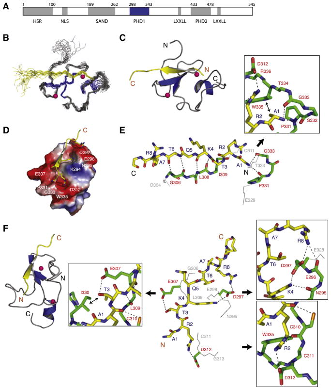
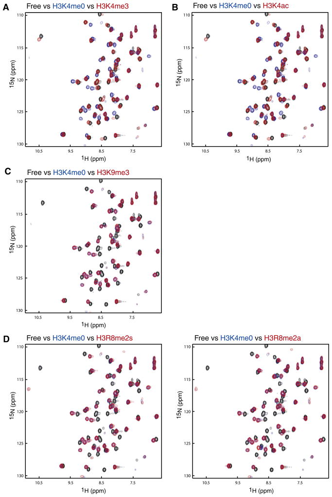
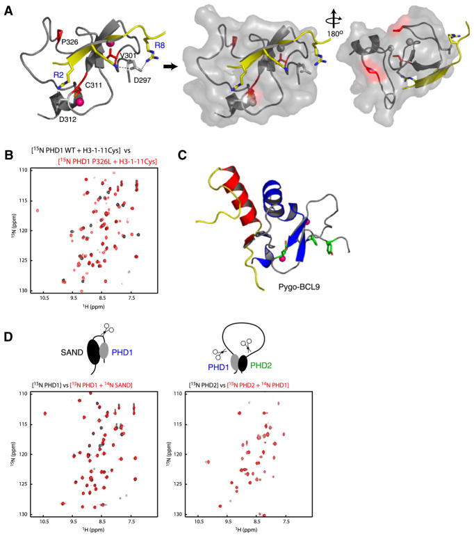
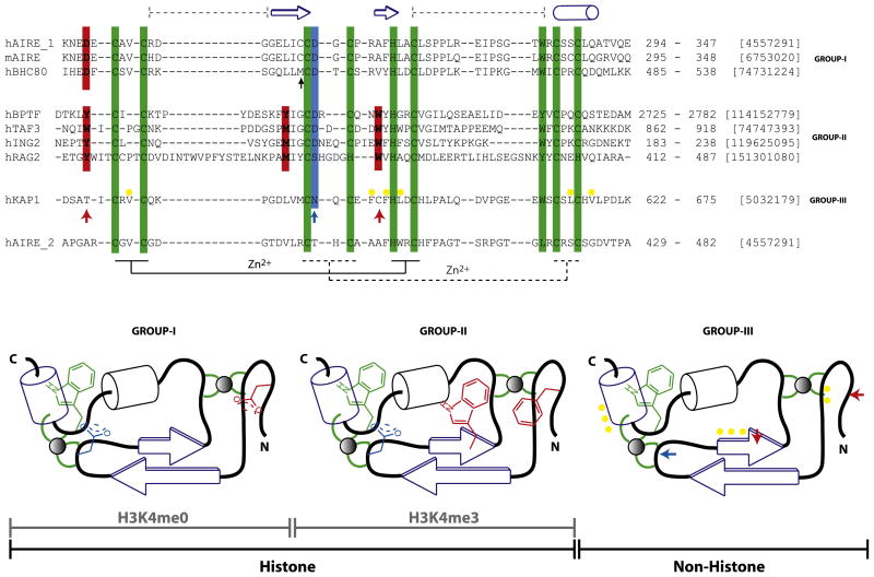
Human autoimmune regulator (AIRE) functions to control thymic expression of tissue-specific antigens via sequence-specific histone H3 recognition by its plant homeodomain (PHD) finger. Mutations in the AIRE PHD finger have been linked to autoimmune polyendocrinopathy-candidiasis-ectodermal dystrophy (APECED). Here we report the three-dimensional solution structure of the first PHD finger of human AIRE bound to a histone H3 peptide. The structure reveals a detailed network of interactions between the protein and the amino-terminal residues of histone H3, and particularly key electrostatic interactions of a conserved aspartic acid 297 in AIRE with the unmodified lysine 4 of histone H3 (H3K4). NMR binding study with H3 peptides carrying known posttranslational modifications flanking H3K4 confirms that transcriptional regulation by AIRE through its interactions with histone H3 is confined to the first N-terminal eight residues in H3. Our study offers a molecular explanation for the APECED mutations and helps define a subclass of the PHD finger family proteins that recognize histone H3 in a sequence-specific manner.


Figures

Figure 1
Figure 1. Three-Dimensional Solution Structure of the AIRE-PHD1/H3K4me0 Peptide Complex
(A) Schematic representation of the functional domains in the human AIRE protein. Grey boxes represent HSR (homogenously staining region), PHD (plant homeodomain), and SAND (Sp100, AIRE-1, NucP41/P75, and Drosophila DEAF-1). PHD and SAND domain boundaries are based on Pfam HMM (Bateman et al., 1999) and remaining segments are based on Meloni et al. (2008). AIRE-PHD1 studied here is shown in blue. (B) Backbone atoms (N, Cα, and C′) of the 20 superposed NMR structures of the AIRE-PHD1 where protein and peptide are gray and yellow, respectively (left). (C) Ribbon representation of the complex (middle) highlights the secondary structural elements (protein, blue; peptide, yellow). Pink spheres represent Zn atoms. Only a single representation of Zn atoms of the lowest energy structure is shown in the ensemble for clarity. (D) Electrostatic potential (isocontour value of ±70 kT/e) surface representation of the AIRE-PHD1 bound to the H3K4me0 peptide (yellow). (E) Backbone protein-peptide interactions with inset showing the H3A1 interacting neighborhood. The peptide and protein residues are color coded by atom type with carbon atoms in yellow and green, respectively. The orientation of the peptide is the same as that in (C). (F) Key protein-peptide side-chain interactions with insets respectively highlighting R2, K4, and T3 neighborhood and their surface grooves. The nonpolar nonbonded interacting atoms are labeled with ↔. The peptide orientation in the stick representation is depicted as in the ribbon diagram on left.
Figure 2
Figure 2. Peptide Binding Studies
Peptide binding studied between AIRE-PHD1 and N-terminal H3 peptides by NMR. Comparison of two-dimensional 1H-15N HSQC spectra of AIRE-PHD1 between its free form (black) and that in presence of a peptide derived from N-terminal H3 residues 1–11 with or without a known post-translational modification: (A) H3K4me0 (blue) versus H3K4me3 (red); (B) H3K4ac (red); (C) H3K9me3 (red); (D) H3R8me2a (red), left panel, and H3R8me2s (red), right panel. The concentration of the protein was 0.5 mM, and the molar ratio of the protein to peptide was kept at 1:5 for all NMR binding studies.
Figure 3
Figure 3. APECED Mutants
(A) Location of the mutant residues (red) on the protein (gray)-peptide (yellow) complex structure in ribbons (left) and transparent surface representation (middle, right). The pink Zn atoms in left panel are shown in gray in surface representation for clarity. (B) Comparison of two-dimensional 1H-15N HSQC spectra of AIRE-PHD1 wild-type with that of P326L mutant in presence of a peptide derived from N-terminal H3 residues 1–11 (black, wild-type; red, P326L mutant). (C) Nonhistone interacting surface of Pygo-PHD (gray) in complex with the HD1 domain of BCL9 (yellow/red) (left). The aromatic cage residues of Pygo-PHD involved in H3K4me2 are green. (D) Two-dimensional 1H-15N HSQC spectra assessing AIRE’s interdomain interaction at 1:1 molar ratio with 0.2 mM respective labeled proteins. The 15N-labeled PHD1 and PHD2 are labeled blue and green, respectively.
Figure 4
Figure 4. Classification of the PHD Finger Family
Sequence features of structurally characterized three distinct subclasses of the PHD finger family with respect to ligand binding specificity, i.e., H3K4me0 (group I), H3K4me3 (group II), and nonhistone binding (group III). The recognition of H3K4me3 takes place by embracement of the trimethyl groups by characteristically positioned aromatic cage residues, whereas that of K4me0 is due to an ion pair formed with a distinct N-terminal Asp residue. The characteristic histone-peptide interacting positions are in red, and the Zn-chelating residues (the first and second tetrads are connected by regular and dotted lines, respectively) and conserved C-terminal aromatic residue characteristic of the entire PHD family are in green. In either of the H3 interacting PHD fingers, H3R2 often interacts with Asp/Glu (blue). These “red” and “blue” positions are absent in KAP1-PHD finger (bottom) indicated by ↑ that binds the adjacent bromodomain’s ZA helix by patch of nonpolar residues (yellow). Topology diagrams (bottom; based on Aravind et al. [2006]), not drawn to scale, highlight these features for clarity. The domain boundaries and gi numbers are indicated in the alignments. The secondary structural elements of the AIRE-PHD1 are indicated above the sequence. The black ↑ indicates position where similar interactions involving protein side chain is observed in AIRE and BHC80 PHD fingers. The sequence of AIRE-PHD2 is shown below the alignment to show its grouping with group III as non-H3 binder.

References

    1. Aasland R, Gibson TJ, Stewart AF. The PHD finger: implications for chromatin-mediated transcriptional regulation. Trends Biochem Sci. 1995;20:56–59. - PubMed
    1. Anderson MS, Venanzi ES, Klein L, Chen Z, Berzins SP, Turley SJ, von Boehmer H, Bronson R, Dierich A, Benoist C, Mathis D. Projection of an immunological self shadow within the thymus by the aire protein. Science. 2002;298:1395–1401. - PubMed
    1. Aravind L, Iyer L. Provenance of SET-domain histone methyl-transferases through duplication of a simple structural unit. Cell Cycle. 2003;2:369–376. - PubMed
    1. Aravind L, Iyer LM, Koonin EV. Comparative genomics and structural biology of the molecular innovations of eukaryotes. Curr Opin Struct Biol. 2006;16:409–419. - PubMed
    1. Bateman A, Birney E, Durbin R, Eddy SR, Finn RD, Sonnhammer EL. Pfam 3.1: 1313 multiple alignments and profile HMMs match the majority of proteins. Nucleic Acids Res. 1999;27:260–262. - PMC - PubMed

Publication types

Associated data